Herunterladen
Kostenlose voll funktionsfähige 14-Tage-Testversion enthalten
DISSECTION MASTER
Reale menschliche anatomie.
Das Studium der komplexen menschlichen Anatomie in virtueller Realität ist einfacher, schneller und genauer als mit herkömmlichen Methoden auf einem flachen Bildschirm oder in Büchern. Gehen Sie um die Sektionen herum, greifen Sie den Körper mit den Händen, vergrößern Sie ihn auf jede Größe und tauchen Sie in die kleinsten anatomischen Strukturen in VR ein.
Der menschliche Körper wird in zehn hochauflösenden Schichten dargestellt. Sie können von den äußeren Muskeln zu den inneren Organen navigieren und tausende anatomische Strukturen bis ins kleinste Detail studieren und lehren.
Mit Dissection Master XR kann die menschliche Anatomie schneller und besser als je zuvor visualisiert, studiert und verstanden werden. Außerdem können Sie Ihre eigenen Lektionen und Simulationen in VR erstellen. Nehmen Sie Ihre Lektionen auf, um sie in VR abzuspielen, und erstellen Sie eine wachsende Bibliothek Ihrer Anatomielektionen.
Dissection Master XR führt zu einem besseren Verständnis der menschlichen Anatomie und Physiologie und schafft eine neue Generation hochqualifizierter medizinischer Fachkräfte.
Beginnen Sie jetzt mit dem Unterrichten der Anatomie mit Dissection Master XR!
Erleben Sie den menschlichen Körper und seine Anatomie in einem vollständig immersiven holografischen Raum. Willkommen bei Dissection Master XR, Ihrem Atlas der menschlichen Anatomie in VR.
Der gesamte menschliche Körper - von Kopf bis Fuß - wird in hervorragender Qualität in zehn immersiven und vollständig annotierten Schichten präsentiert.
Mehr als 3000 anatomische Strukturen sind benannt, kommentiert und mit zusätzlichen Informationen verknüpft.
Vergrößern Sie den menschlichen Körper, untersuchen und studieren Sie ihn aus jedem Winkel und jeder Perspektive. Vergleichen Sie ihn mit medizinischen Bildern und den anatomischen Daten von Zygote mit den anderen Medicalholodeck-Apps.
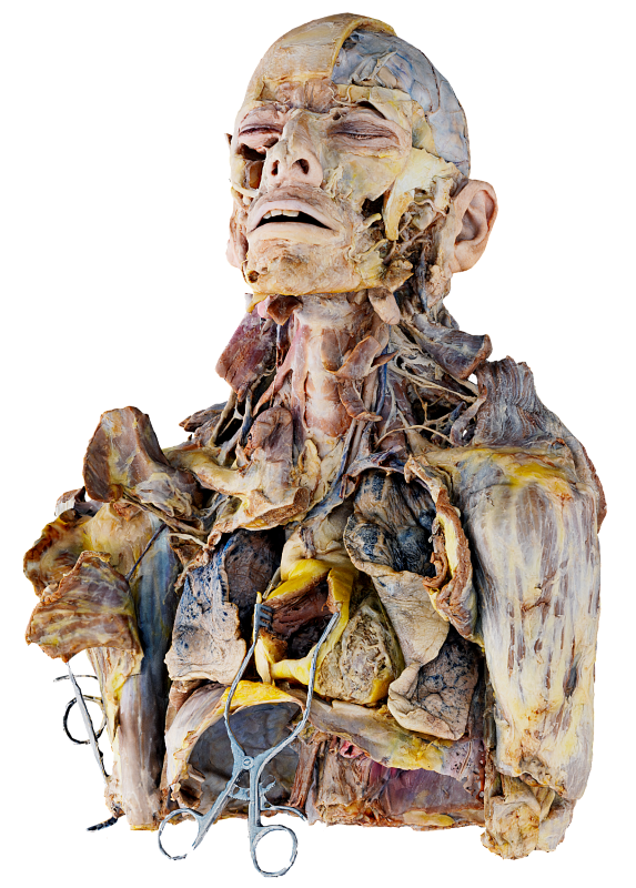
Die Präparationen werden sorgfältig von einem führenden Spezialisten erstellt.
Echte menschliche Präparationen bilden die Grundlage des Atlas. Nach der Präparation wurde der Körper sorgfältig mit modernster Technologie digitalisiert.
Lehren Sie menschliche Anatomie in Ihrer Abteilung und ortsunabhängig in virtuellen Klassenzimmern im Metaversum.
Nehmen Sie Anatomielektionen auf, entwickeln Sie Ihre Inhalte und speichern und teilen Sie Ihr Wissen mit Nutzern weltweit.
App-Inhalt
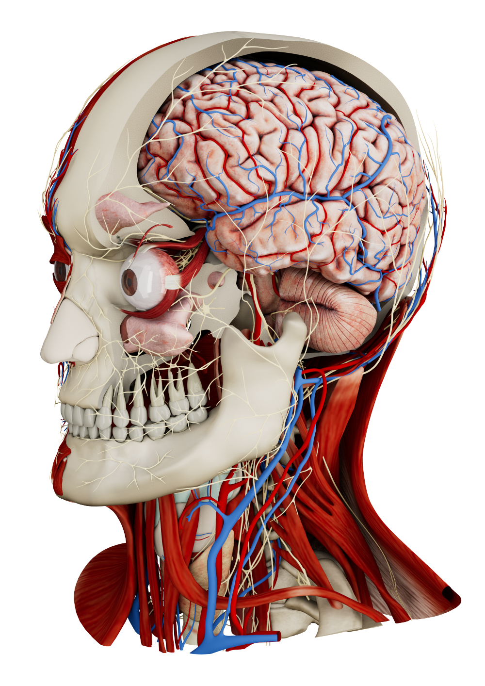
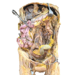
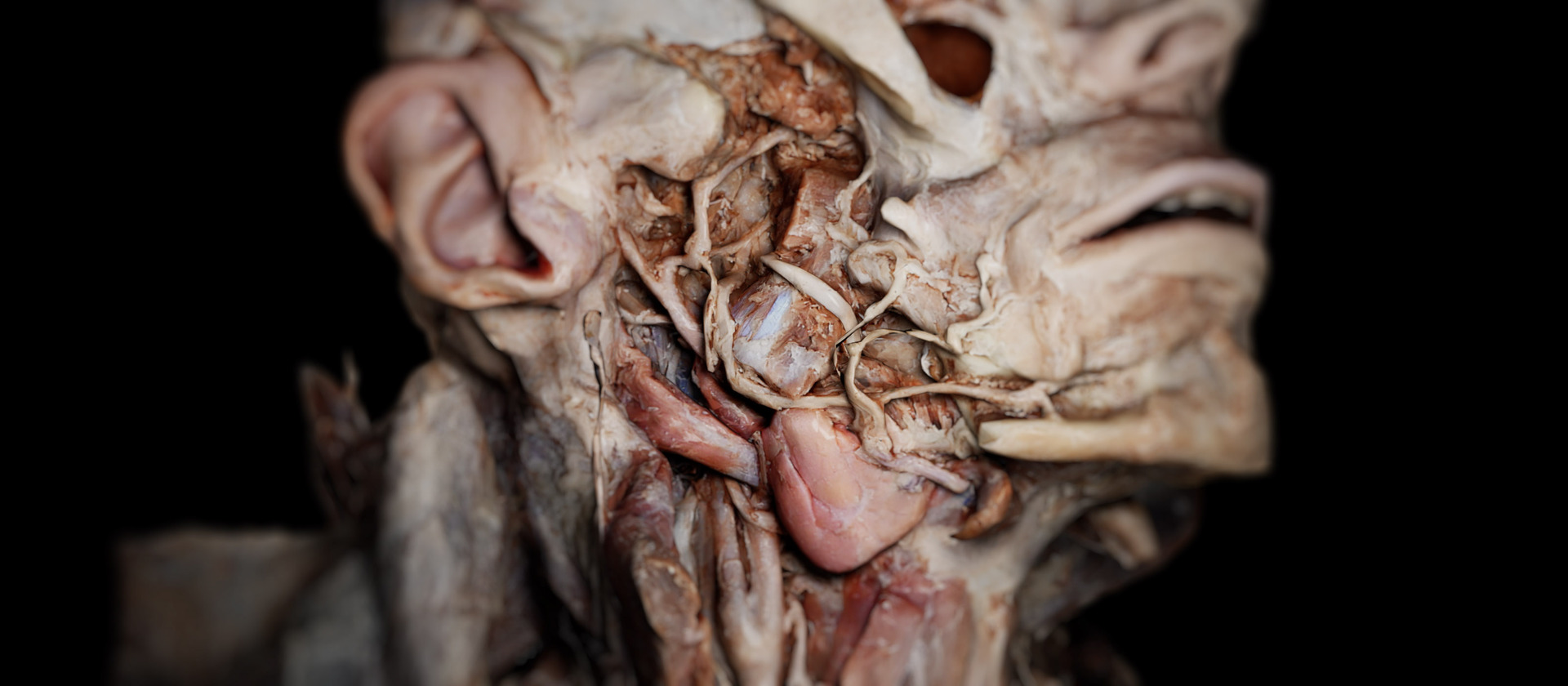
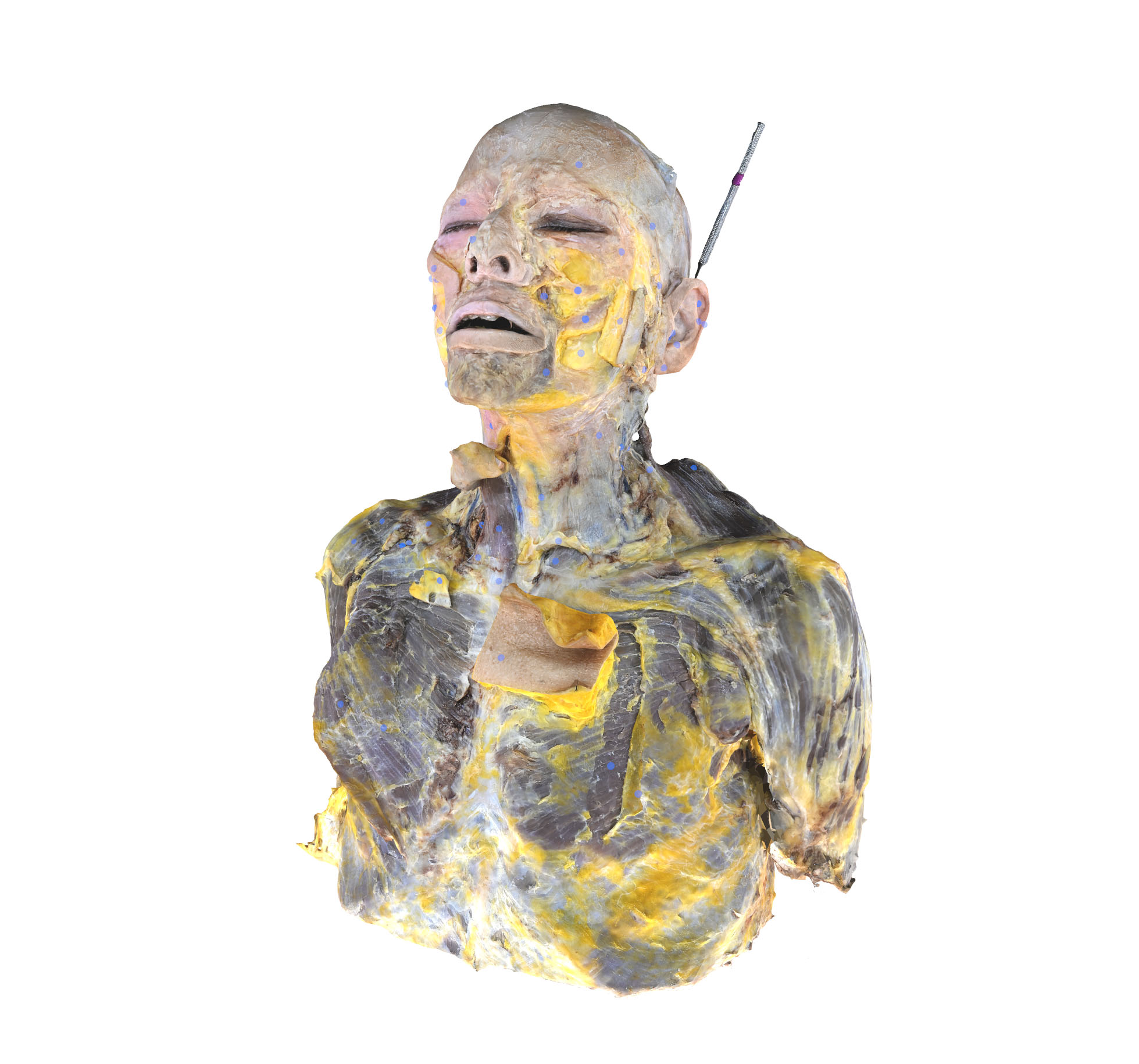
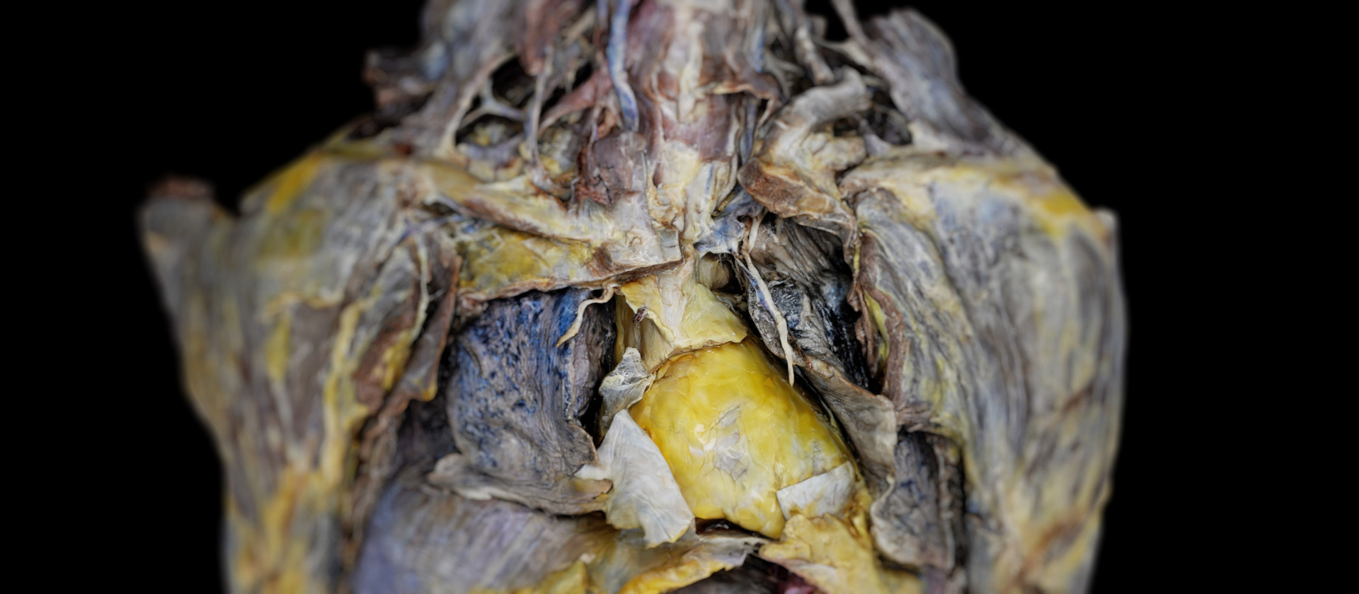
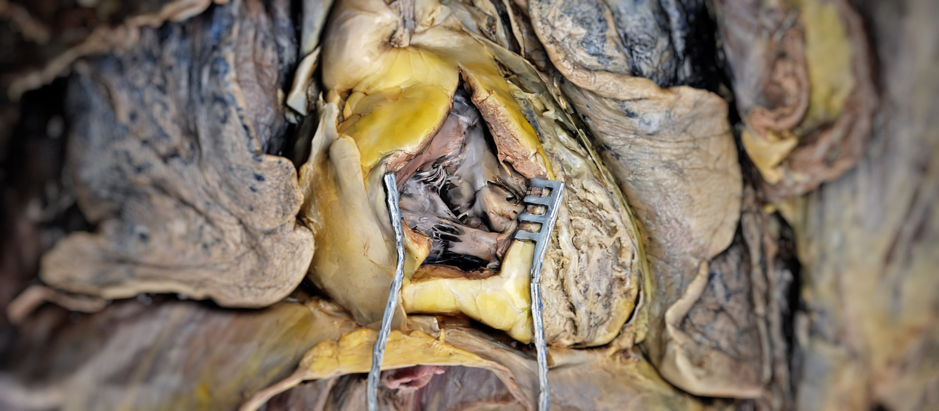
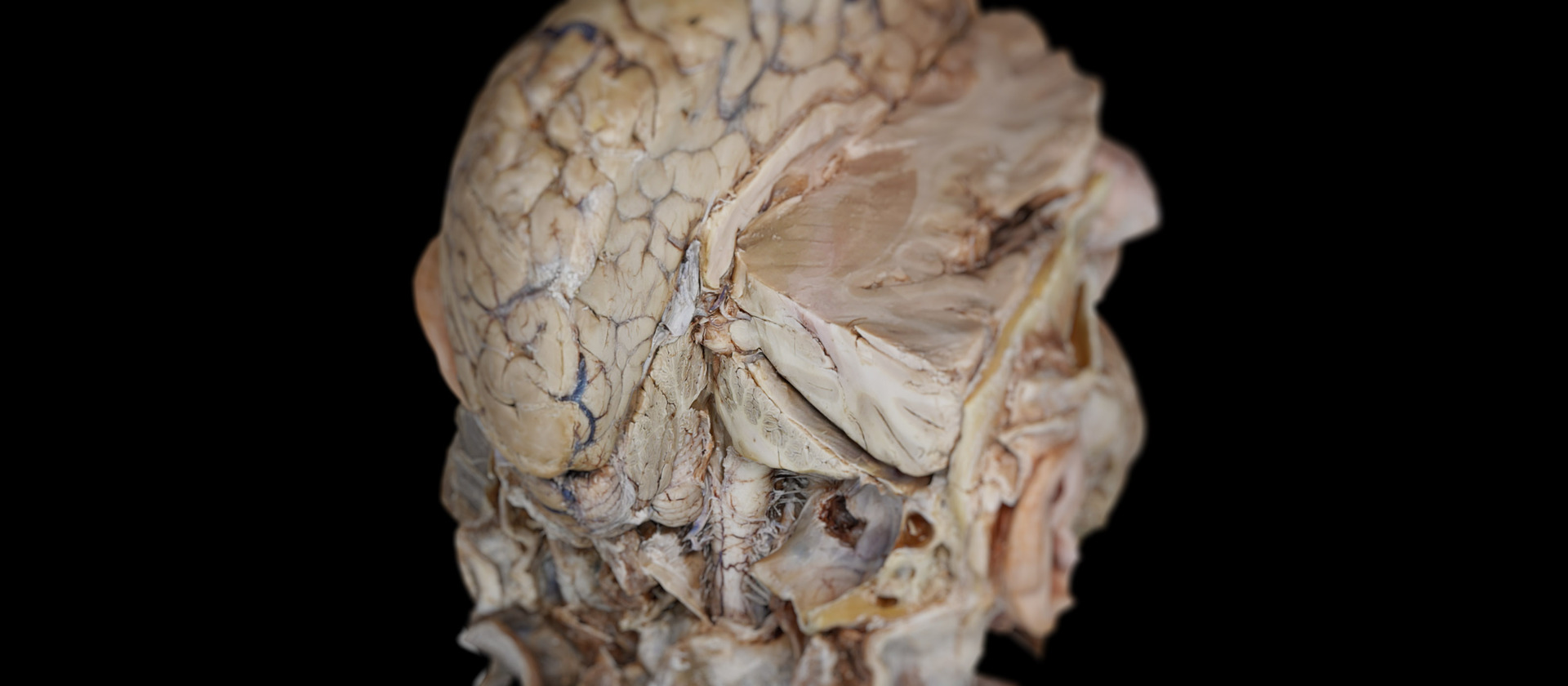
 Kopf und Thorax
Erkunden Sie zehn Präparationsschichten von Kopf und Thorax mit über 3500 beschrifteten Strukturen – von Haut und mimischer Muskulatur bis zu den tiefsten Organen und Gefäßen. Untersuchen Sie mehr als 150 Muskeln, Gefäße und Nerven des Halses zusammen mit Halswirbeln, Bandscheiben und Rückenmark.
Studieren Sie die Brusthöhle mit Herz, Koronararterien sowie großen systemischen und pulmonalen Gefäßen. Sehen Sie Lungen, Trachea, Ösophagus und den Thoraxrahmen.
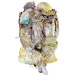
 Weibliches Abdomen
Untersuchen Sie das weibliche Abdomen und Becken in sieben Präparationsschichten mit über 3000 beschrifteten Strukturen. Von der Bauchwand mit ihren Muskeln und Gefäßen bis zum Verdauungssystem mit Magen, Leber, Pankreas, Darm, Milz, Nieren und Nebennieren.
Große Gefäße wie Aorta abdominalis und Vena cava inferior werden mit ihren Ästen gezeigt, neben der Beckenhöhle mit Blase, Rektum und Fortpflanzungsorganen. Beckenboden, Knochen, Gelenke, Bänder und Bindegewebe sowie Plexus lumbalis und sacralis erscheinen schichtweise.
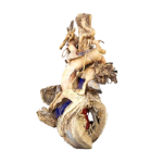
 Männliches Abdomen
Erkunden Sie vierzehn anatomische Schichten des männlichen Abdomens und Beckens. Untersuchen Sie den Rumpf von den Bauchmuskeln und der Bauchwand bis zu den tiefen kardiovaskulären und nervalen Systemen. Die innere Anatomie zeigt das Verdauungssystem mit Speiseröhre, Leber und Gallenblase sowie Dünn- und Dickdarm. In tieferen Schichten werden Nieren, Nierenvenen und Harnblase sichtbar, zusammen mit den männlichen Fortpflanzungsorganen.
 Herz
Herz
Das Herzpräparationsmodell zeigt die Außenfläche mit den großen Gefäßen, Vorhöfen und Ventrikeln. Die innere Anatomie präsentiert Klappen, Septen, Trabekel, Papillarmuskeln und Chordae tendineae. Koronararterien und -venen werden mit animiertem arteriellen und venösen Blutfluss dargestellt.
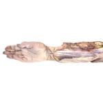 Arm
Arm
Erkunden Sie die obere Extremität in zehn Präparationsschichten mit fast 1000 beschrifteten Strukturen. Von Haut, Venen und Muskelkonturen bis zu den Flexor- und Extensorkompartimenten des Unterarms. Thenar- und Hypothenarmuskeln, Interossei, Lumbricales und Sehnenscheiden führen zum vollständigen Skelettgerüst mit Gelenken, Bändern, Gefäßen und Nerven im gesamten Arm.
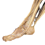 Bein
Bein
Über 1600 beschriftete Strukturen des Beins werden in acht Präparationsschichten gezeigt. Von Haut, Venen und großen Muskelgruppen bis zu den tiefsten Kompartimenten. Schicht für Schicht erscheinen die Gesäßmuskeln, die Adduktoren des Oberschenkels sowie die Flexor- und Extensorenkompartimente des Unterschenkels.
Der Fuß zeigt die intrinsischen Muskeln, einschließlich Interossei und Lumbricales, mit plantaren Strukturen. Das Skelett – Femur, Tibia, Fibula und Patella – mit Bändern und Gelenken; das arterielle System reicht von der Femoralarterie bis zu tibialen Ästen.
Erkunden Sie zehn Präparationsschichten von Kopf und Thorax mit über 3500 beschrifteten Strukturen – von Haut und mimischer Muskulatur bis zu den tiefsten Organen und Gefäßen. Untersuchen Sie mehr als 150 Muskeln, Gefäße und Nerven des Halses zusammen mit Halswirbeln, Bandscheiben und Rückenmark.
Studieren Sie die Brusthöhle mit Herz, Koronararterien sowie großen systemischen und pulmonalen Gefäßen. Sehen Sie Lungen, Trachea, Ösophagus und den Thoraxrahmen.
Das Verständnis der menschlichen Anatomie erfordert ausgezeichnete dreidimensionale Visualisierungen. Dissection Master XR ist Ihr interaktiver Anatomieatlas in virtueller Realität und bietet die weltweit höchste Auflösung und detaillierteste anatomische Präparationen in einem vollständig immersiven holografischen Raum.
Unterrichten und studieren Sie menschliche Anatomie in VR, verbessern Sie die Qualität Ihrer Ausbildung und fördern Sie die Lernfähigkeiten Ihrer Studierenden.
Verbinden Sie sich mit Ihren Studierenden und unterrichten Sie Anatomie in virtuellen Klassenzimmern. Ortsunabhängiges Lehren eröffnet neue Möglichkeiten für die universitäre Bildung, internationale Zusammenarbeit und globales Lernen.
Sparen Sie Zeit und steigern Sie Ihren Erfolg. Das Lernen von 3D-Strukturen in einer immersiven 3D-Umgebung ist einfacher als auf flachen Bildschirmen oder aus Büchern.
Erstellen Sie Ihre Anatomielektionen, Simulationen und Inhalte in VR und machen Sie sie Nutzern weltweit zugänglich.
Nutzen Sie Dissection Master XR und die Daten und Apps von Medicalholodeck, um menschliche Anatomie in virtuellen Klassenzimmern zu unterrichten. Fügen Sie Ihre Medien hinzu und erstellen Sie Lektionen in virtueller Realität. Bauen Sie Ihre Bibliothek mit Bildungsinhalten und Simulationen auf und teilen Sie sie mit Studierenden und Nutzern weltweit im Metaversum.
Unterrichten Sie Anatomie in VR, fügen Sie Ihre DICOM-, 3D- und 2D-Dateien hinzu und erstellen Sie Lektionen, Simulationen und Inhalte für die Wiedergabe in VR. Nutzen Sie Medicalholodeck, um Ihr bestehendes Präparationslabor zu verbessern und Ihr Simulationslabor auf die nächste Stufe zu bringen.
Studieren Sie die menschliche Anatomie in virtueller Realität mit Dissection Master XR, Anatomy Master XR und Medical Imaging XR. Alle Anwendungen können gleichzeitig genutzt werden und bieten Studierenden außergewöhnliche Einblicke in menschliche Präparationen, anatomische Modelle und medizinische Bildgebung. Nutzer bestätigen, dass das Lernen in virtueller Realität Zeit gespart hat und sie in ihrer medizinischen Karriere erfolgreicher gemacht hat.