Explore the abdomen and pelvis in layered detail, from the anterior abdominal wall to deep pelvic structures. This VR model highlights the inguinal area, femoral triangle, gluteal region, anal fossa, and cauda equina. Examine abdominal viscera, peritoneal membranes, retroperitoneal organs, pelvic arteries, kidney structures, the pelvic floor, and lumbar and sacral plexus nerves, an invaluable tool for studying muscles, vessels, and neurovascular anatomy in context.
Price
Free
Duration
00:04:14
Languages
English (Original)
Difficulty
File type
RXR
Author
Evan Goldman, Ph.D. Director of Anatomy, Associate Professor, Penn State University College of Medicine
Publication date
June 2023
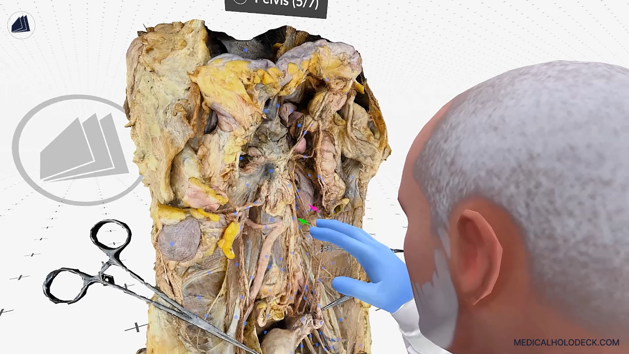
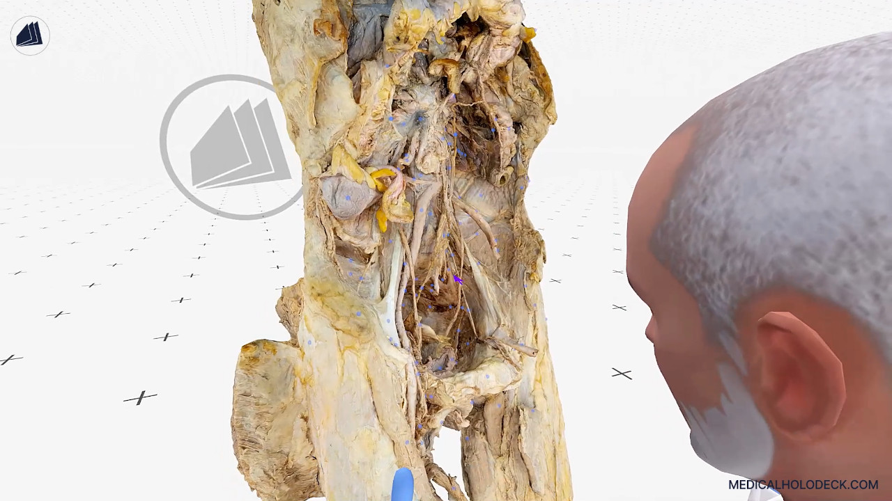
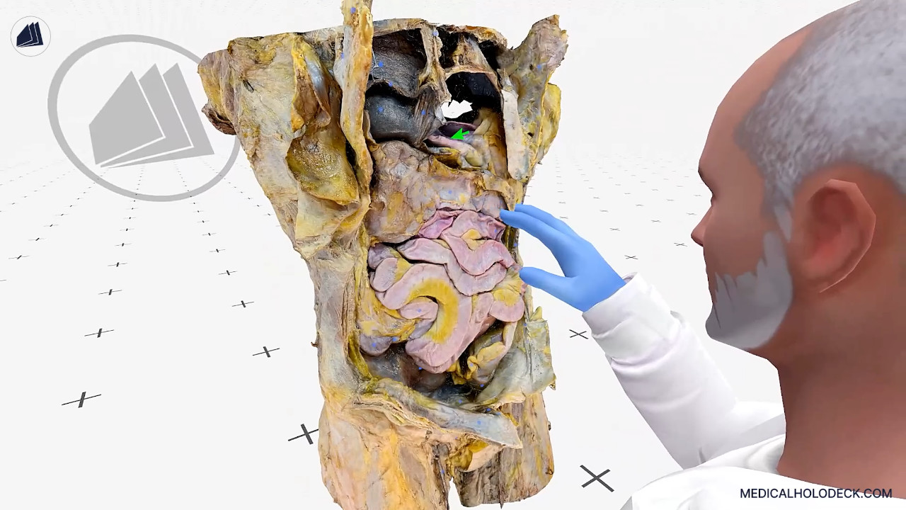
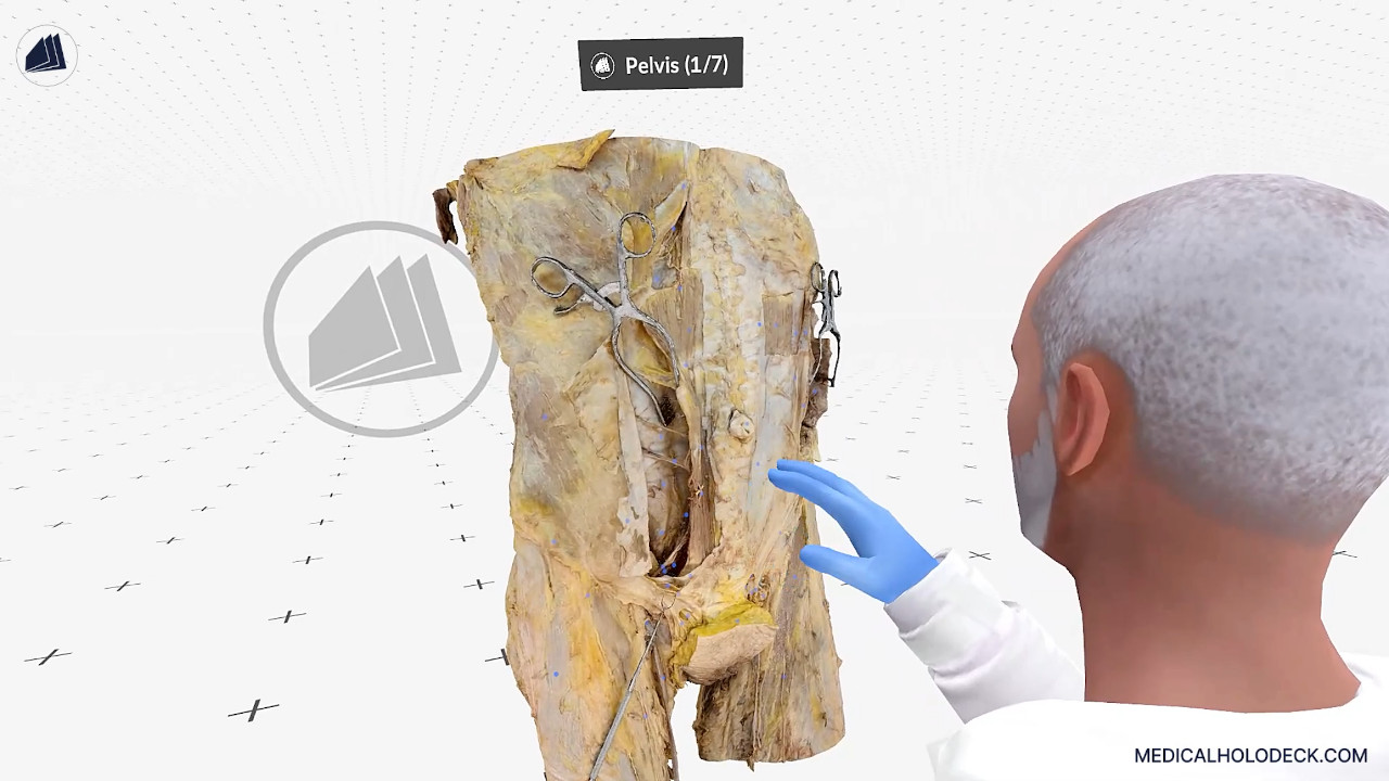
In this VR lesson, Evan describes and demonstrates how the abdomen and pelvis model reveals a wide range of anatomical regions, including the inguinal area, femoral triangle, external female genitalia, gluteal region, anal fossa, and cauda equina. He begins with the anterior abdominal wall, showing its muscles, vessels such as the inferior epigastric artery, and associated membranes.
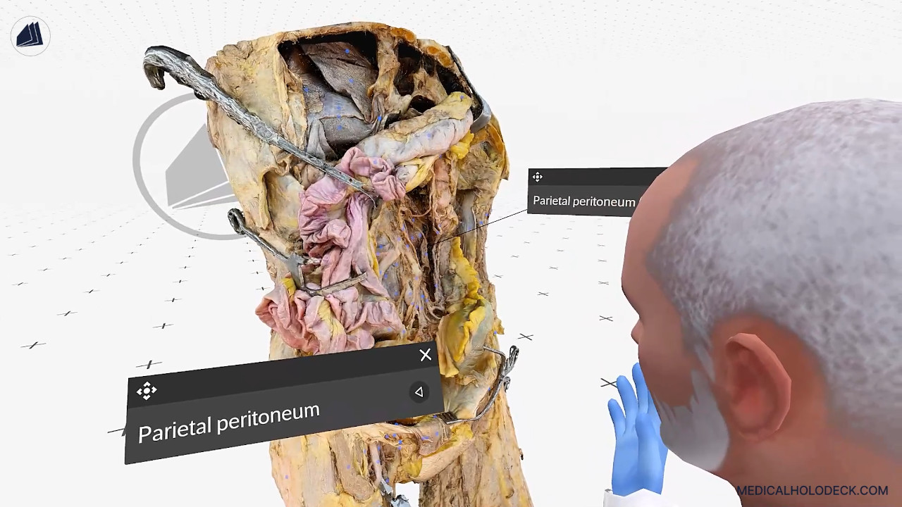
Deeper layers reveal abdominal viscera , the liver, stomach, and intestines of the foregut, midgut, and hindgut, along with the parietal peritoneum and retroperitoneal or subperitoneal structures like the uterus and bladder.