Anatomy Master enhances the understanding of human anatomy more effectively and efficiently compared to traditional methods like screen-based learning or textbooks. This application offers a dynamic learning experience where users can interact with anatomical data by walking around, enlarging, dissecting, and making cross-sectional views of the models, allowing for observation and study from any angle and perspective.
Anatomy Master facilitates a rapid and enhanced understanding of the human body, its organs, and physiology. Utilizing this tool leads to a deeper comprehension of human anatomy, resulting in more proficient medical students, nurses, and healthcare professionals.
With Medicalholodeck, educators have the capability to create their own lessons and simulations in VR. This feature allows for the recording of teachings for VR replay, enabling the development of a comprehensive library of anatomy lessons.
Anatomy Master can be effectively utilized in virtual classrooms or for individual student self-study, enhancing the learning curve and contributing to the improved education of future medical professionals through VR technology.
Download
Free fully functional 14-day trial included
For iPad and
iPhone
i
For iPhone and iPad
View medical imaging and 3D anatomy in augmented reality on
mobile devices.
App Store
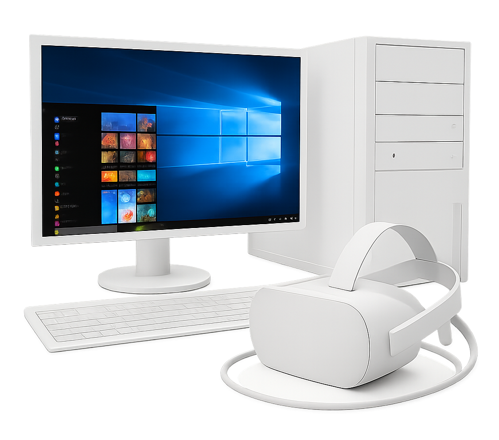
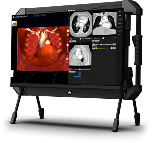
For tethered
PC-VR
i
For headsets connected to a PC
View large CT/MRI scans, segment anatomy with AI, and explore
3D anatomy in virtual reality.
Performance depends on your computer’s hardware capabilities.
Download
Steam
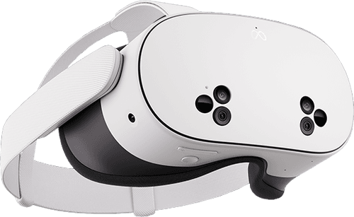
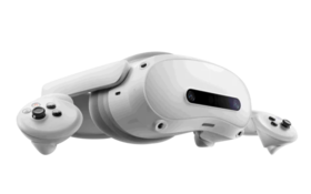
For Pico 4
Ultra
i
For Pico 4 Ultra
View standard-sized CT/MRI scans, segment anatomy with AI,
and explore 3D anatomy in virtual reality.
Limited performance may occur with medical imaging.
For full performance, use PC-VR.
Pico Store
For glasses-free 3D and 2D
screens
i
For 3D and 2D screens
Compatible with both 3D naked eye displays
and standard 2D screens on Windows computers.
Naked eye 3D screens provide an immersive,
true spatial view without the need for VR headsets.
Request Beta access
Key features
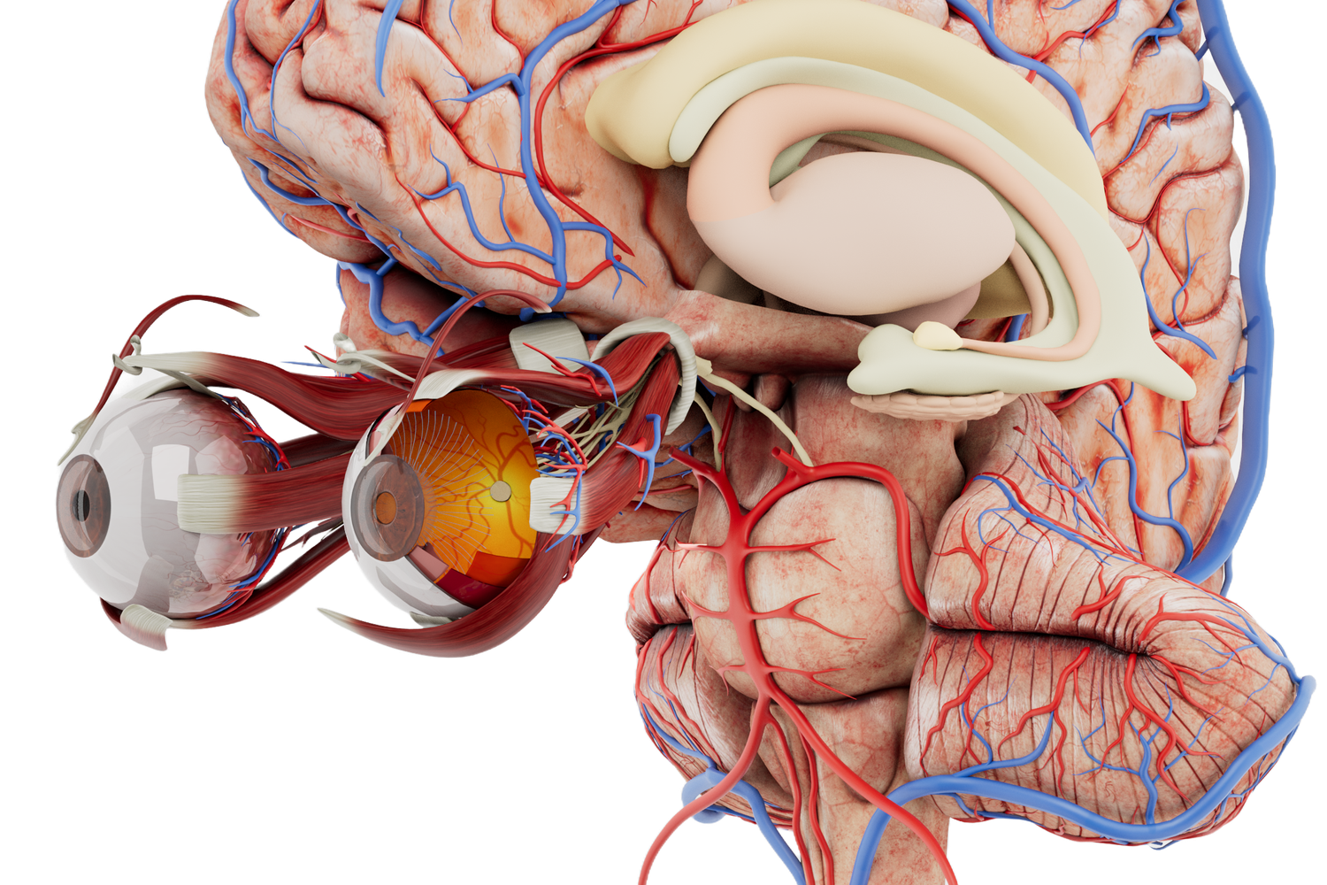
Three-dimensional VR anatomy
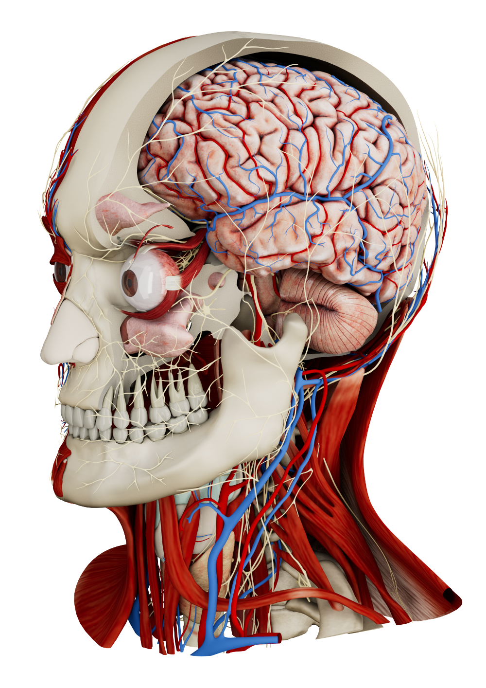
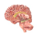
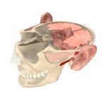
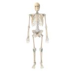
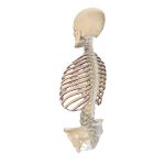
Anatomy Master XR presents high-quality, three-dimensional human anatomy in VR, ideal for teaching and studying the human body in immersive virtual classrooms.
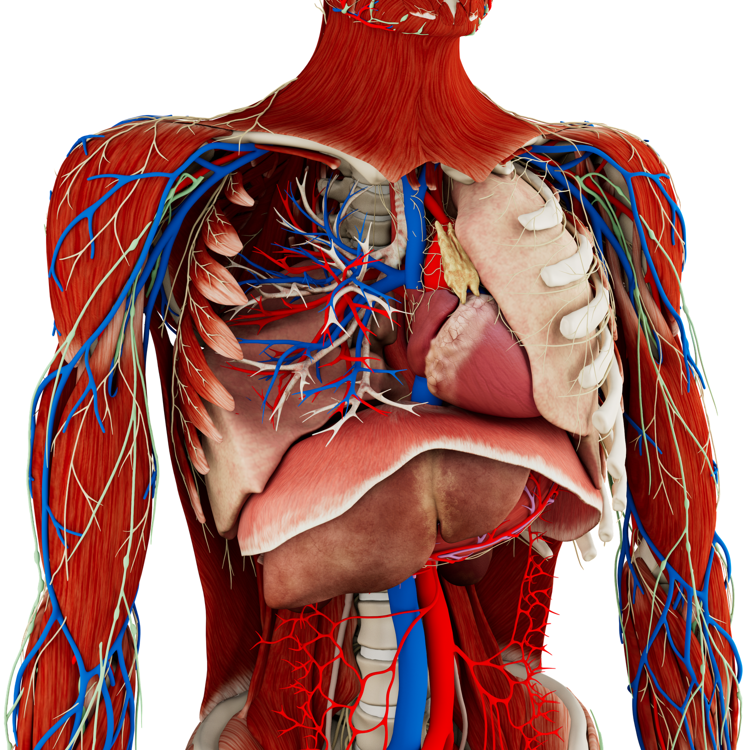
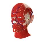
Accuracy
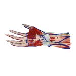
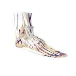
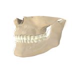
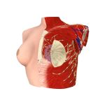
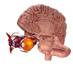
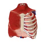
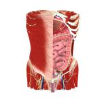
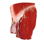
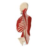
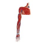
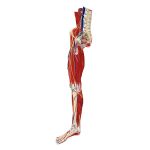
The application includes precise male and female anatomy models, developed based on real patient data, ensuring accuracy and reliability in virtual reality. Medicalholodeck has developed the models in collaboration with leading experts to ensure a professional-grade educational tool.
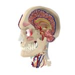
Annotated models
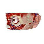
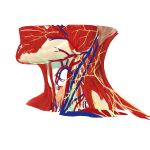
Over 2000 anatomical structures and organs are meticulously annotated, supporting detailed study and effective teaching in virtual classroom environments and self-study.
Immersive experience
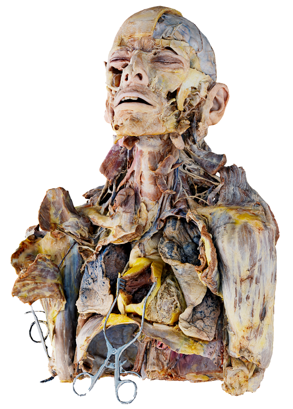
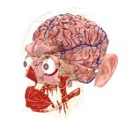
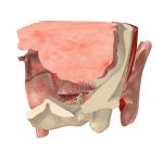
Users can magnify the anatomical models, study them from any perspective, and compare them side-by-side with medical imaging and dissections from our comprehensive anatomy atlas in a fully immersive workspace.
High-quality modeling
The 3D models are based on CT and MRI data, with textures and structures carefully crafted to accurately represent real-life organs.
Collaborative learning
The platform enables teaching human anatomy in various settings - from virtual classrooms and VR labs to location-independent global access.
Record, replay, share
Educators can record anatomy classes, create and store lessons, and share their content globally, allowing users to replay and learn individually.
Advantages
3D anatomy models in virtual reality
High-quality three-dimensional visualization is crucial for understanding human anatomy, particularly in the initial stages of medical education. Virtual reality offers an immersive and detailed perspective that enhances learning.
Enhanced knowledge acquisition
Utilizing VR, students can explore the human body's organs and anatomical structures in a more interactive and engaging manner. This approach elevates the quality of education and student performance, fostering the development of the next generation of medical professionals in a virtual reality environment.
Comprehensive anatomical understanding
Offering complete three-dimensional anatomical insights in VR significantly improves students' grasp of human anatomy. This method is more efficient and effective compared to traditional learning resources, facilitating faster and deeper comprehension.
Teaching in virtual classrooms
Virtual classrooms in the metaverse enable location-independent teaching, creating opportunities for international collaboration and learning. Medicalholodeck's comprehensive medical XR applications provide a platform for teaching, studying, and learning anatomy in an innovative way.
Record, store, share in VR
Educators can easily create, store, and share their anatomy lessons and simulations with a simple click, making these resources readily accessible to users in the metaverse. This feature enhances the distribution and accessibility of educational content in the field of medical education.

 icon.jpg)