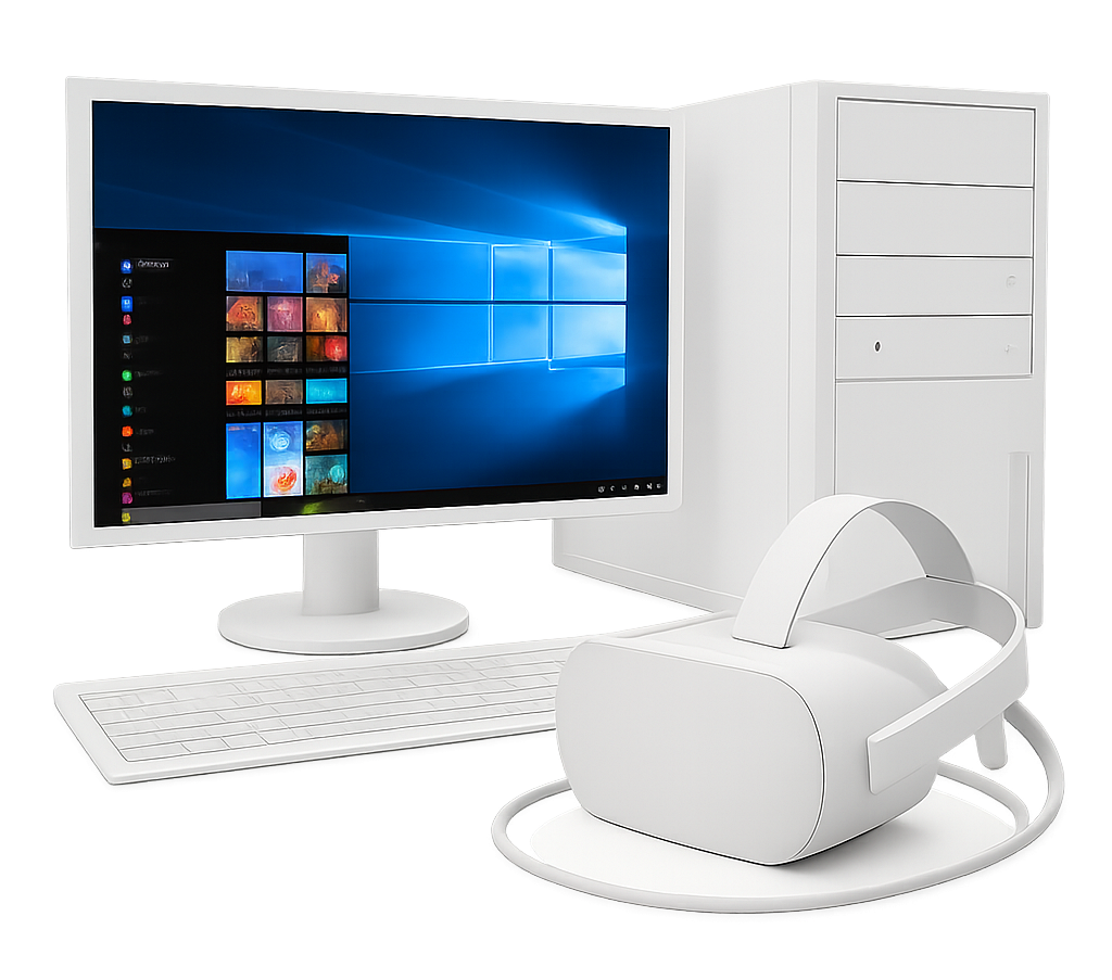
Download
Free fully functional 14-day trial included
DISSECTION MASTER
Real human anatomy.
Studying complex human anatomy in virtual reality is easier, faster, and more accurate than with conventional methods on a flat screen or in books. Walk around the dissections, grab the body with your hands, enlarge them to any size, and dive into the smallest anatomical structures in VR.
The human body is displayed in ten hi-res layers. You can browse from the outer muscles to the inner organs and study and teach thousands of anatomical structures in utmost detail.
With Dissection Master XR, human anatomy can be visualized, studied, and understood faster and better than ever before. And you can create your own lessons and simulations in VR. Record your teachings for replay in VR, and build a growing library of your anatomy lessons.
Dissection Master XR leads to an improved understanding of human anatomy and physiology and creates a new generation of highly skilled medical professionals.
Start teaching anatomy with Dissection Master XR now!
Experience the human body and its anatomy in a fully immersive holographic space. Welcome to Dissection Master XR, your human anatomy atlas in VR.
The whole human body - from head to toe - is offered in superb quality in ten immersive and fully annotated layers.
More than 3000 anatomical structures are named, annotated, and linked to additional information.
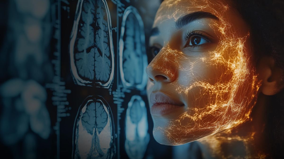
Enlarge the human body, and examine and study it from any angle and perspective. And compare it with medical imaging and Zygote's anatomy data using the other Medicalholodeck apps.
The dissections are carefully created by a leading specialist.
Real human dissections form the basis of the atlas. After dissection, the body was carefully digitized using the latest state-of-the-art technology.
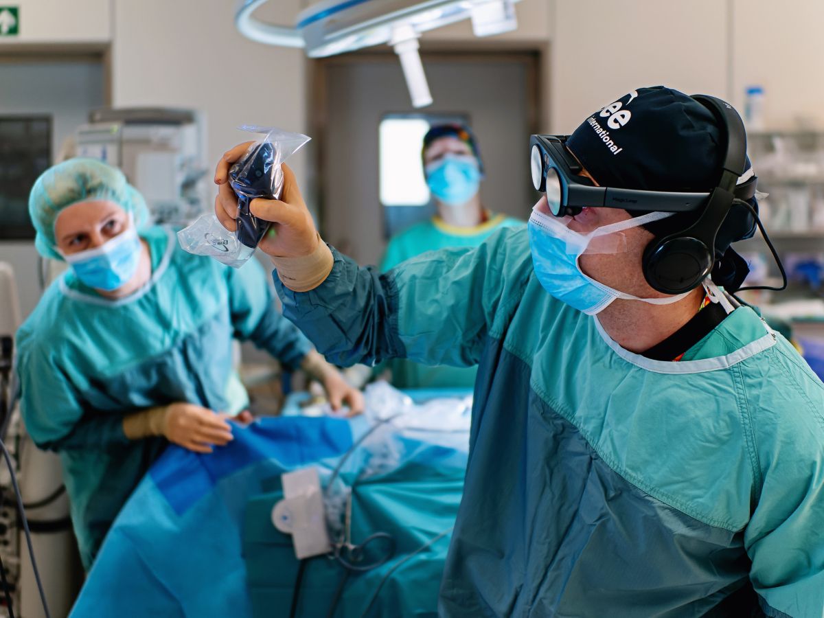
Teach human anatomy at your department and location-independent in virtual classrooms in the metaverse.
Record anatomy lessons, develop your content, and store and share your know-how with users worldwide.
App content
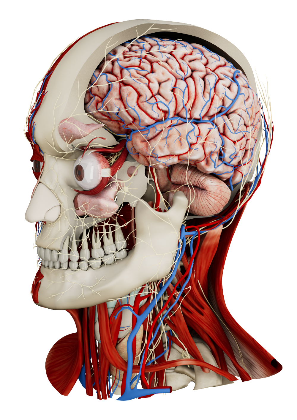
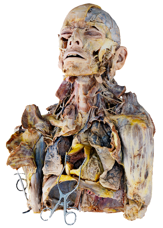
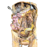
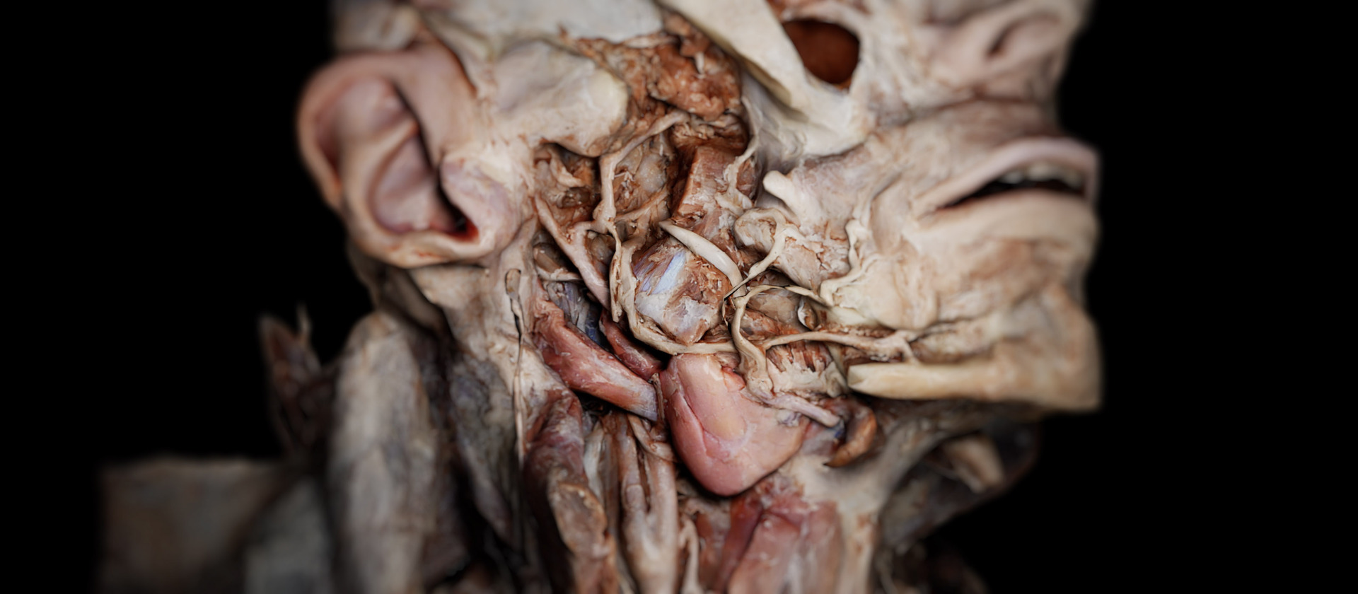
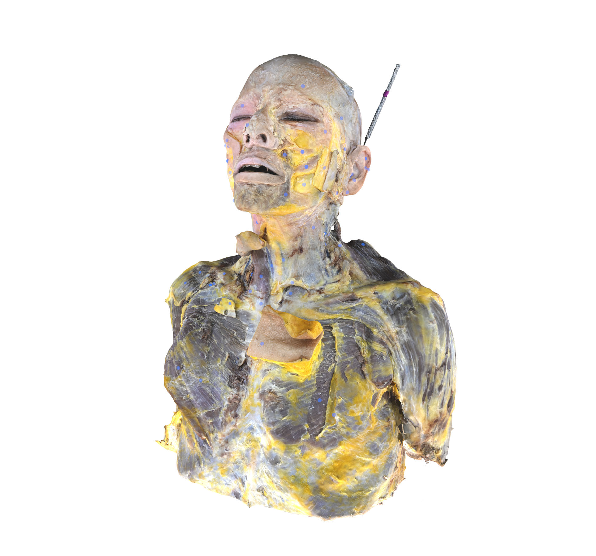
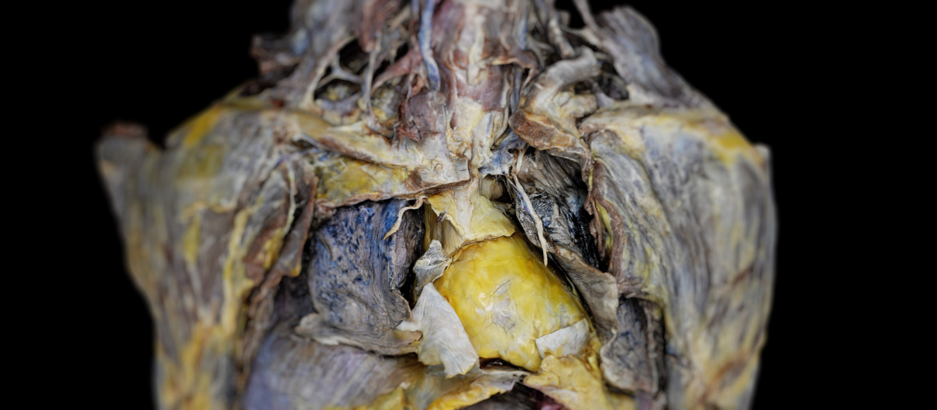
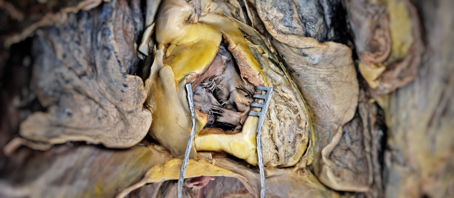
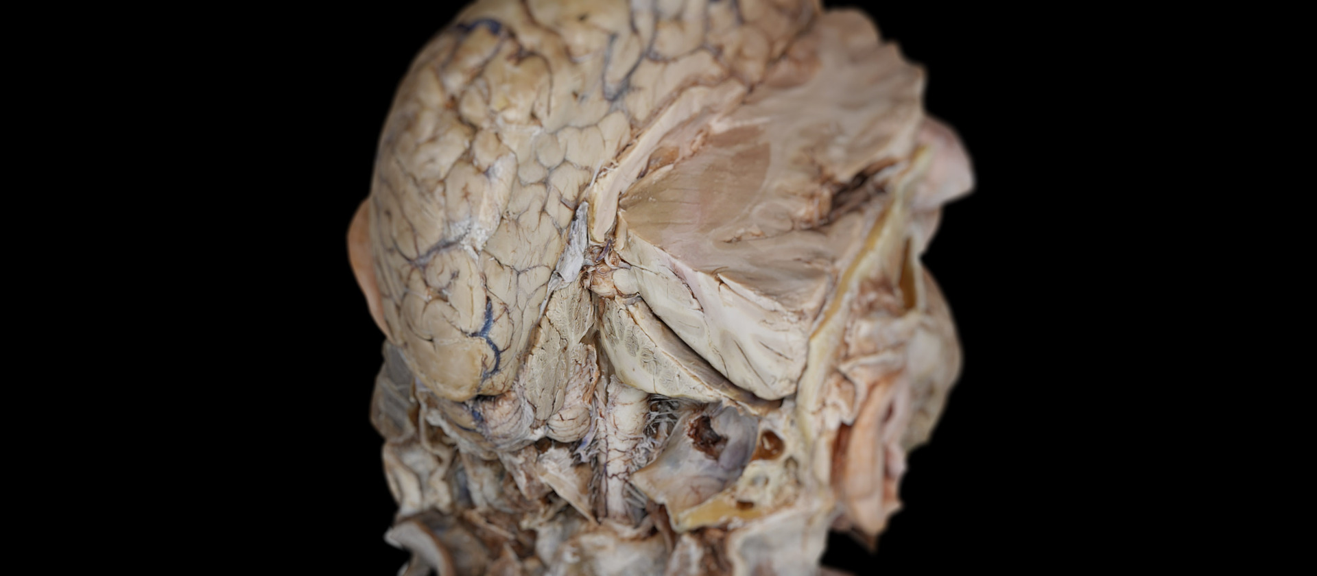
 Head and thorax
Explore ten dissection layers of the Head and thorax with more than 3500 annotated structures, from the skin and facial muscles to the deepest organs and vessels. Examine more than 150 muscles, vessels, and nerves of the neck together with the cervical vertebrae, intervertebral discs, and spinal cord.
Study the thoracic cavity with the heart, coronary arteries, and great systemic and pulmonary vessels. View the lungs, trachea, esophagus, and rib cage.
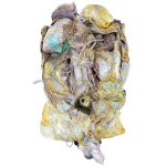
 Female abdomen
Examine the female abdomen and pelvis in seven dissection layers, featuring over 3000 annotated structures. From the abdominal wall with its muscles and vessels to the digestive system with the stomach, liver, pancreas, intestines, spleen, kidneys, and adrenal glands.
Major vessels such as the abdominal aorta and inferior vena cava are presented with their branches, alongside the pelvic cavity with bladder, rectum, and reproductive organs. The pelvic floor, bones, joints, ligaments, and connective tissues are revealed layer by layer, together with the lumbar and sacral plexuses.
 Male abdomen
Explore fourteen anatomical layers of the male abdomen and pelvis. Examine the trunk from abdominal muscles and walls through the deep cardiovascular and nervous systems. The inner anatomy features the digestive system, including the esophagus, liver, and gallbladder, along with the small and large intestines. Deeper layers reveal kidneys, renal veins, and urinary bladder, together with male reproductive organs.
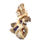 Heart
Heart
The heart dissection model shows the external surface with the great vessels, atria, and ventricles. The inner anatomy presents the valves, septa, trabeculae, papillary muscles, and chordae tendineae. The coronary arteries and veins are shown with arterial and venous blood flow illustrated in motion.
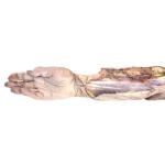 Arm
Arm
Explore the upper limb in ten dissection layers with nearly 1000 annotated structures. From skin, veins, and muscle contours to the forearm’s flexor and extensor compartments. Thenar and hypothenar groups, interossei, lumbricals, and tendon sheaths, culminating in the full skeletal framework with joints, ligaments, vessels, and nerves traced throughout the entire limb.
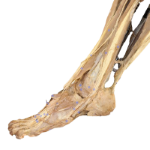 Leg
Leg
Over 1600 annotated structures of the leg are presented across eight dissection layers. From the skin, veins, and major muscle groups to the deepest compartments. Layer by layer, the module reveals the gluteal muscles, thigh adductors, and the flexor and extensor compartments of the leg.
The foot shows the intrinsic muscles, including interossei and lumbricals, with plantar structures. The skeleton: femur, tibia, fibula, and patella with ligaments and joints, while the arterial system extends from the femoral to tibial branches.
Explore ten dissection layers of the Head and thorax with more than 3500 annotated structures, from the skin and facial muscles to the deepest organs and vessels. Examine more than 150 muscles, vessels, and nerves of the neck together with the cervical vertebrae, intervertebral discs, and spinal cord.
Study the thoracic cavity with the heart, coronary arteries, and great systemic and pulmonary vessels. View the lungs, trachea, esophagus, and rib cage.
Understanding human anatomy requires excellent three-dimensional visualizations. Dissection Master XR is your interactive anatomy atlas in virtual reality, offering the world's highest resolution and most detailed anatomical dissections in a fully immersive, holographic space.
Teach and study human anatomy in VR, improve your education's quality, and enhance your students' learning skills.
Connect with your students and teach anatomy in virtual classrooms. Location-independent teaching opens new possibilities for university education, international collaboration, and global learning.
Save time and increase your success. Learning 3D structures in an immersive 3D environment is easier than from flat screens or books.
Create your anatomy lessons, simulations, and content in VR, and make it accessible to users worldwide.
Use Dissection Master XR and Medicalholodeck's data and apps to teach human anatomy in virtual classrooms. Add your media, and create lessons in virtual reality. Build your library of educational content and simulations, and share it with students and users worldwide in the metaverse.
Teach anatomy in VR, add your DICOM, 3D, and 2D files, and create teachings, lessons, and simulations for replay in VR. Use Medicalholodeck to enhance your existing dissection lab and bring your simulation lab to the next level.
Study human anatomy in virtual reality with Dissection Master XR, Anatomy Master XR, and Medical Imaging XR. All applications can be used simultaneously, giving students extraordinary insight into human dissections, anatomy models, and medical imaging. Users confirm that studying in virtual reality saved time and made them more successful in their medical careers.