Enhancing Sentinel Lymph Node Biopsy in Endometrial Cancer Using Augmented and Mixed Reality. Cureus 17(1): e77599.
https://doi.org/10.7759/cureus.77599Sentinel lymph node (SLN) biopsy
Sentinel lymph node (SLN) biopsy is a minimally invasive technique used to assess lymph node status in endometrial cancer. By identifying and removing only the first draining lymph nodes, it provides accurate staging while significantly reducing the morbidity associated with full lymphadenectomy, such as lymphedema, nerve injury, and vascular complications.
SLN detection typically involves the combined use of the radioisotope (RI) method and the near-infrared fluorescence imaging method. While the RI method is effective for detecting SLNs, it does not provide visual feedback during surgery, necessitating the use of indocyanine green. The ICG method also has limitations, such as the detection of multiple SLNs and the difficulty of identifying SLNs in patients with obesity.
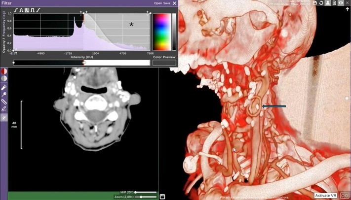
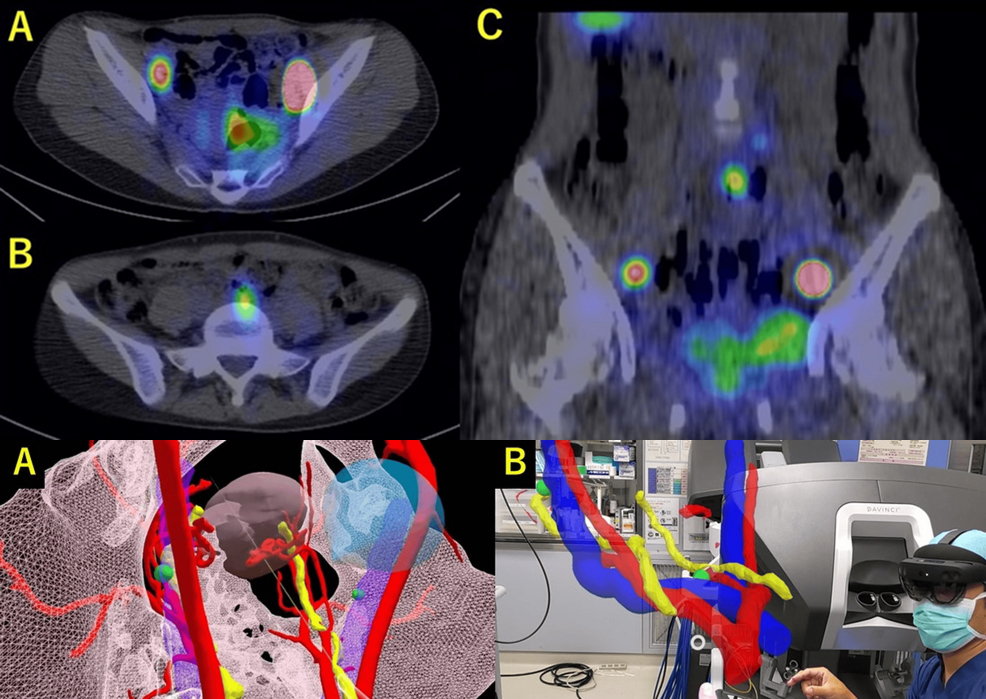
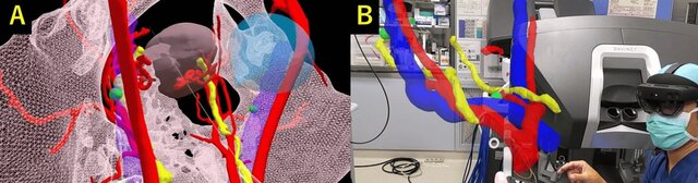
Sentinel lymph node identified by SPECT/CT.
Integrating AR and MR
Augmented reality and mixed reality are increasingly used in surgical procedures. In endometrial cancer biopsies, they have been applied for preoperative visualization of sentinel lymph nodes and their anatomical relationships, as well as for intraoperative navigation. Mixed Reality was integrated into surgical procedures through the use of head-mounted displays (HMDs). Holographic projections provided surgeons precise guidance during surgery, improving lymph node identification and minimizing invasiveness. This allowed the team to better understand the spatial relationships between the SLNs, blood vessels, and ureters, increasing the precision of SLN biopsy.
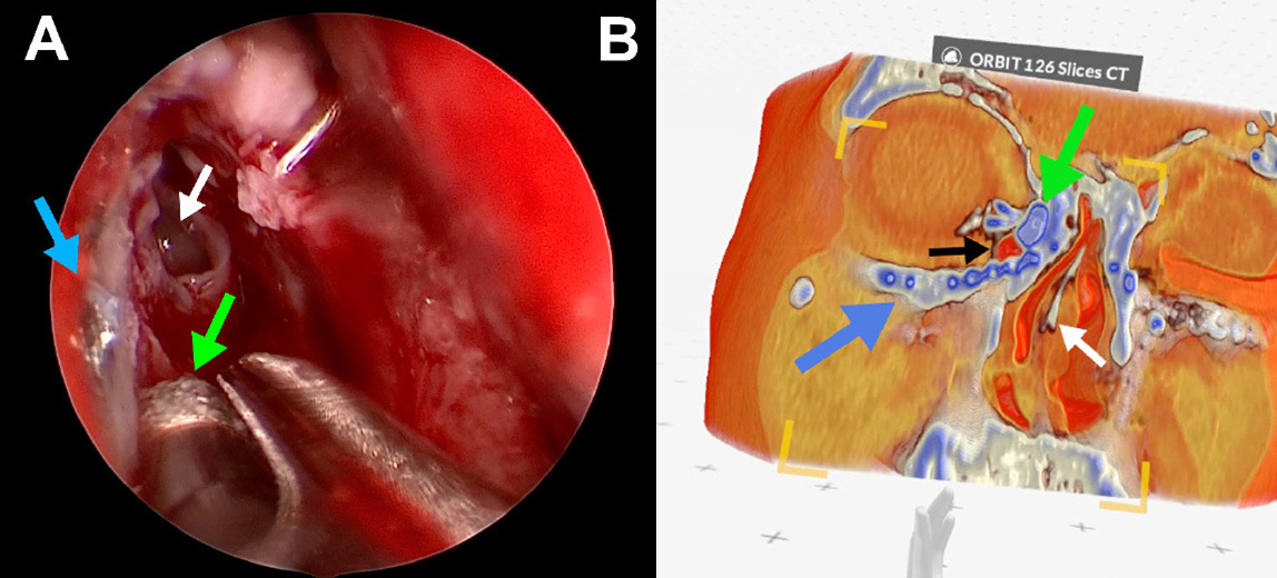
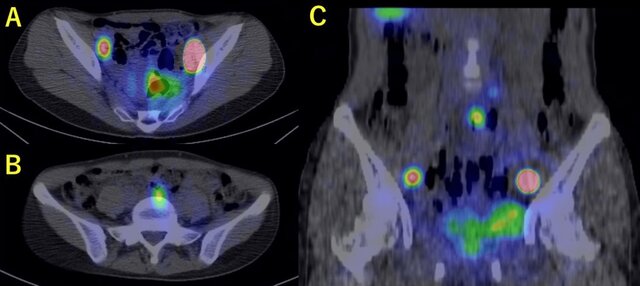
Holograms showing the positional relationship between blood vessels, ureters, and sentinel lymph nodes. The green spheres represent the sentinel lymph nodes.
Implementing AR and MR allowed the tailored visualization of 3D anatomical reconstructions based on individual patient imaging.This approach simplified lymph node identification and supported selective SLN removal while preserving surrounding structures.
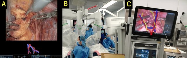
Robot-assisted laparoscopic sentinel lymph node biopsy under hologram guidance.
Holography-guided AR is particularly well suited for SLN procedures, because lymph nodes and blood vessels are fixed anatomical structures that remain stable despite changes in body position. An assistant wearing a see-through headset can overlay holographic images onto the endoscopic camera view, enabling a clear understanding of the lymph nodes targeted by the surgeon.
This shared visualization improves surgical accuracy and coordination between the surgeon and assistant, having the potential to reduce complications and shorten operative time.
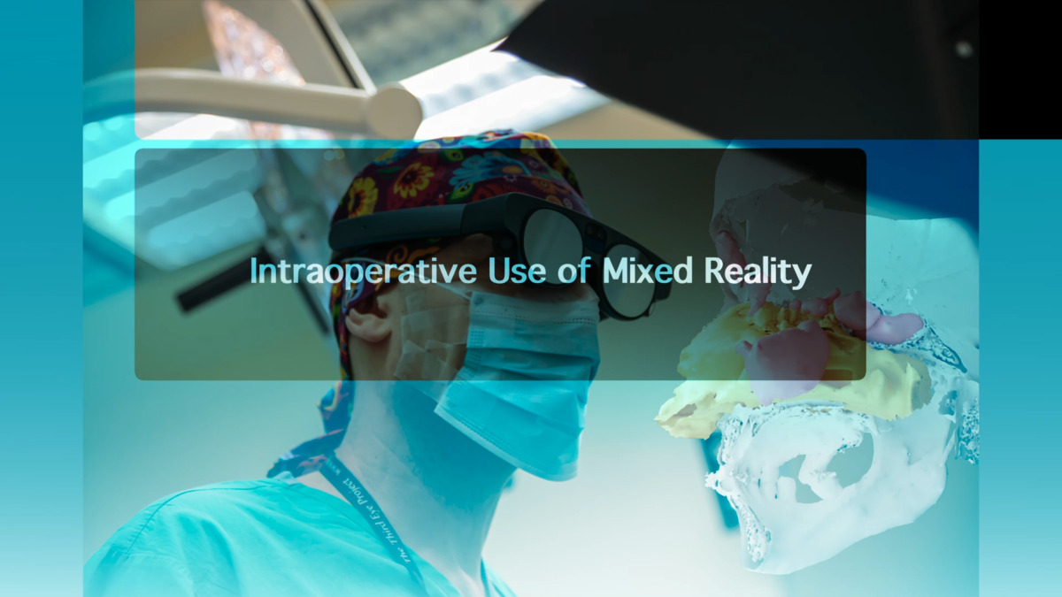
Medicalholodeck for intraoperative navigation
Augmented reality offers a practical solution by projecting three-dimensional anatomical models directly onto the patient. Instead of diverting attention to external monitors or relying solely on intraoperative exploration, surgeons can reference holographic visualizations in real time.
With Medical Imaging XR surgeons can create digital twins based on the patient's imaging data. By overlaying reconstructed anatomical structures directly into the operative environment, it supports intraoperative navigation, spatial orientation, and precise identification of target regions. This enhances surgical planning and execution while improving situational awareness for the entire surgical team.
Future of gynecologic oncology
Holography-assisted SLN biopsy is a valuable tool in the surgical management of endometrial cancer. By improving the surgeon’s spatial awareness, it enables more precise identification and excision of sentinel lymph nodes. This case illustrates the successful application of augmented reality in guiding SLN biopsy, resulting in an effective and minimally invasive procedure.
Further integration of holography-assisted AR may expand its role in gynecologic oncology. As these technologies evolve, they have the potential to improve standardization, reduce operative variability, and support wider adoption of less invasive surgical techniques. Continued clinical evaluation will be essential to validate their impact on outcomes and workflow efficiency.
For more information, contact info@medicalholodeck.com March 2026