Intraoperative Holographic Navigation Using Augmented Reality for Facial Artery and Peroneal Perforator Identification in Microsurgical Mandibular Reconstruction.
https://www.preprints.org/manuscript/202508.0103The fibula free flap has become the global standard for major mandibular reconstruction thanks to its length, stable cortical structure, and dependable dual blood supply. Despite technological progress in virtual surgical planning, surgeons still face the challenge of accurately transferring preoperative planning into the operative field, particularly when tumors displace or obscure normal anatomy. Precise visualization of recipient and donor vessels is critical to ensure flap success and long-term function.
Augmented reality in the operating room
Augmented reality offers a practical solution by projecting three-dimensional anatomical models directly onto the patient. Instead of diverting attention to external monitors or relying solely on intraoperative exploration, surgeons can reference holographic visualizations in real time.
This maintains concentration, improves spatial understanding, and assists in identifying structures such as the facial artery and peroneal perforators. The system merges digital planning with physical anatomy, aiming to reduce uncertainty and increase operative efficiency.
Case overview
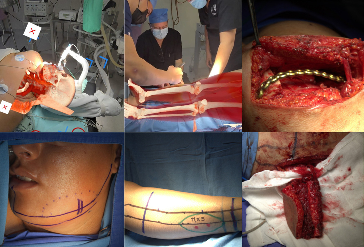
A surgical team in Latin America recently applied low-cost augmented reality navigation in a complex mandibular reconstruction. A 26-year-old woman presented with a large left mandibular osteoblastoma causing progressive swelling and anatomical distortion. CT angiography was used to generate patient-specific 3D models of the mandible, facial vessels, and lower limb vasculature. These datasets were uploaded into a commercially available headset and visualization platform.
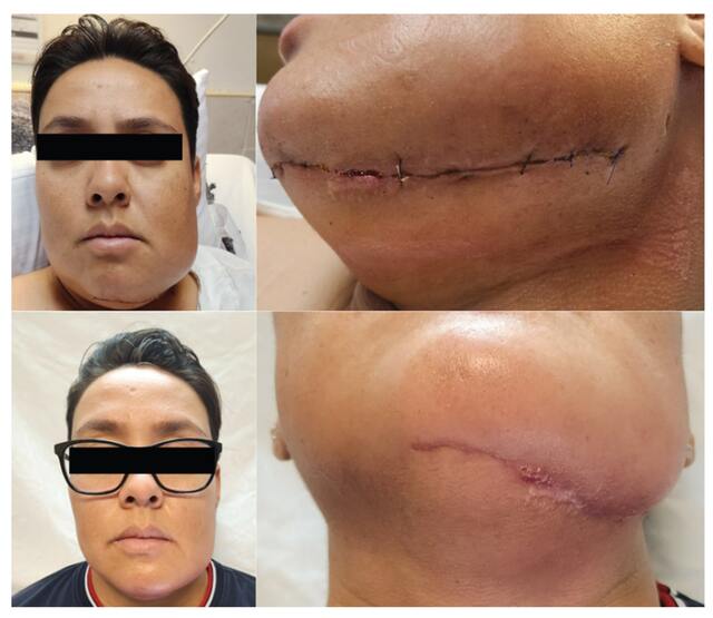
Clinical Follow-up of Postoperative Mandibular Resection. Evolution of facial edema. A comparison is made between the marked facial inflammation at 24 hours postoperatively and the notable improvement with absence of significant inflammation at the 3-month follow-up. Surgical wound healing process. At 24 hours, the wound presents sutures and serous drainage. At 3 months, the incision has healed completely, with only a small granulomatous lesion remaining along the scar line.
Intraoperative AR guidance
During surgery, the primary surgeon wore the head-mounted display to superimpose the hologram over the patient. The tumor mass had displaced the facial artery, but AR guidance enabled clear visualization of the vessel’s new course, supporting safe dissection and preservation.
At the donor site, peroneal perforators were accurately located and marked on the skin using the holographic overlay, minimizing exploration. A 12 cm osteo-fascial fibula flap was harvested, shaped to restore the mandibular contour, and secured with titanium fixation. Microvascular anastomosis to the facial vessels was successfully completed.
Postoperative course and outcome
Recovery proceeded without complications. Oral intake began within 48 hours and advanced to a soft diet by day five. Donor and recipient sites healed satisfactorily, and three-month follow-up demonstrated stable aesthetics, full return to daily activities, and no wound issues.
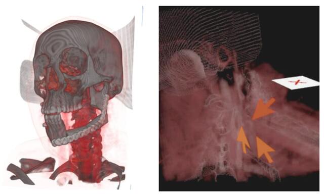
Postoperative Control of Free Fibula Flap Reconstruction. Three-dimensional holographic model demonstrating the postoperative result. The precise fit of the fibular bone flap and the correct positioning of the titanium plate and fixation screws are evident. Assessment of vascular anastomosis patency. The three arrows indicate adequate blood flow at the junction of the flap's peroneal artery with the facial artery.
Benefits of holographic navigation
This case shows that effective digital surgical navigation does not require specialized, high-cost equipment. Consumer-grade augmented reality devices can provide reliable intraoperative guidance at significantly lower expense, making the approach accessible to a wider range of hospitals and surgical services.
The system improved vessel identification, supported the transfer of precise planning to surgery, and maintained surgeon focus within the operative field. It also offers strong value for surgical education, allowing trainees to observe complex anatomical relationships without interrupting workflow or sterility.
Holographic navigation represents a meaningful development in reconstructive microsurgery. By directly integrating virtual planning with the intraoperative view, it narrows the gap between preoperative imaging and surgical execution. As augmented reality platforms continue to evolve, they may become standard tools for reconstruction, surgical planning, and education across multiple specialties.
About Medicalholodeck
Medical Imaging XR allows clinicians to visualize and assess patient data in an immersive spatial environment. Users can interact with patient-specific 3D models in real time, improving anatomical understanding, diagnostic confidence, and the quality of surgical planning.
Medicalholodeck connects securely to hospital systems, offering PACS integration, HIPAA-compliant data handling, and full patient data protection. It runs on VR headsets, PCs, iPads, and iPhones, making it suitable for hospitals, classrooms, and training facilities.
Advanced surgical planning functions are available in Medical Imaging XR PRO FDA. At present, Medicalholodeck is available for educational use only. The platform is in FDA and CE certification, and we expect Medical Imaging XR PRO FDA to enter the U.S. and EU markets soon.
For more information, contact info@medicalholodeck.com November 2025