Virtual reality-assisted preoperative planning for pediatric thoracoscopic segmentectomy: a retrospective study. BMC Pediatrics.
https://doi.org/10.1186/s12887-025-06259-3Improving thoracoscopy with spatial 3D imaging
Thoracotomy has long been a cornerstone in the surgical management of pulmonary conditions. Thoracoscopy offers a tissue-sparing alternative that can significantly improve patient recovery. Compared to open surgery, thoracoscopic procedures are associated with less blood loss, faster recovery, and minimal loss of healthy lung tissue.
Thoracoscopy is technically demanding and requires excellent spatial understanding of complex pulmonary anatomy. Conventional 2D or static 3D imaging is often insufficient for precise preoperative planning.
In contrast, 3D virtual reality models transform standard medical imaging into dynamic, interactive representations, enhancing anatomical comprehension and enabling more accurate and personalized surgical preparation.
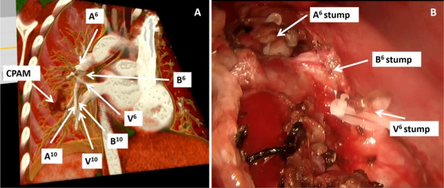
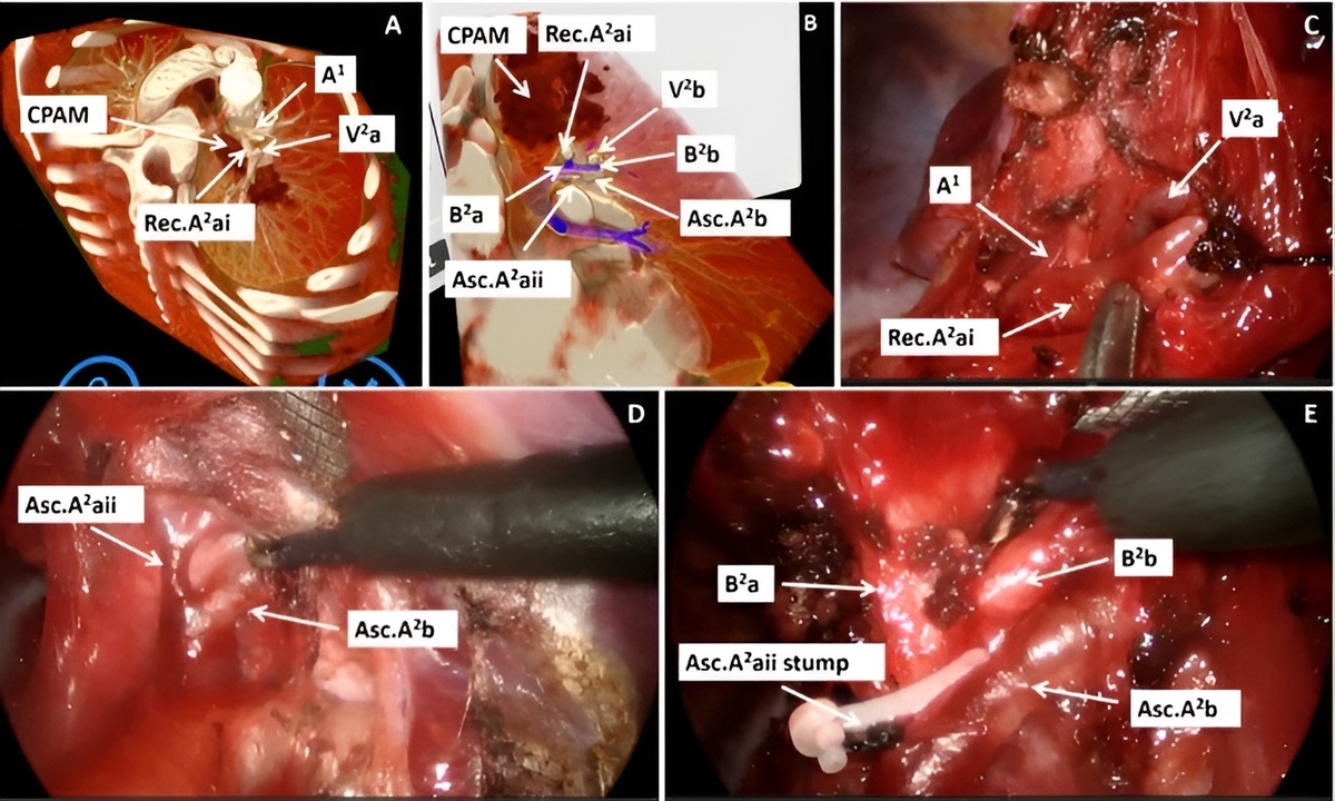
Preoperative Virtual Reality Reconstruction and Corresponding Intraoperative Visualization of Target Vasculature and Bronchial Structures. A Preoperative virtual reality three-dimensional reconstruction planning diagram. B Intraoperative situation. (CPAM: Congenital Pulmonary Airway Malformation; A⁶: Pulmonary Artery to Superior Segment of Lower Lobe; V⁶: Pulmonary Vein from Superior Segment of Lower Lobe; A¹⁰: Posterior basal segmental pulmonary artery; V¹⁰: Posterior Basal Segment Pulmonary Vein; B⁶: Bronchus to Superior Segment of Lower Lobe; B¹⁰: Posterior Basal Segment Bronchus)
Virtual Reality in preoperative planning
The main aim of this study was to evaluate the utility of standalone headsets for preoperative planning in pediatric thoracoscopic segmentectomies. Nineteen pediatric patients who underwent thoracoscopic anatomical segmentectomy were analyzed. Patient-specific three-dimensional models were generated using Medical Imaging XR, based on contrast-enhanced CT data.
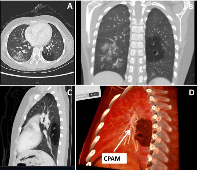
Comparative Visualization of Conventional Computed Tomography and Virtual Reality Reconstruction Using Medicalholodeck Software. A Conventional CT cross-sectional view. B Conventional CT coronal view. C Conventional CT sagittal view. D Virtual reality three-dimensional virtual reconstruction. (CPAM: Congenital Pulmonary Airway Malformation)
Following model reconstruction, the surgical team collaboratively reviewed the spatial relationships between lesions and adjacent critical structures within the VR environment. To validate anatomical fidelity, two independent attending surgeons on associate professor level or higher evaluated all preoperative imaging, focusing on the accuracy of pulmonary arterial branching, vascular calibers, and venous courses.
Based on this preoperative planning, thoracoscopic surgeries were performed with sequential dissection of target vessels and bronchi guided by VR-derived landmarks. The effectiveness of VR reconstruction in facilitating intraoperative navigation was continuously evaluated.
Safer thoracoscopic resections
All 19 procedures were successfully completed thoracoscopically, with no conversions to thoracotomy. Preoperative VR reconstructions provided superior anatomical delineation compared to conventional CT, with two independent assessors confirming enhanced visualization of segmental bronchial branching and spatial relationships with pulmonary vasculature.
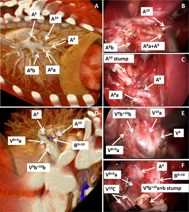
Preoperative VR reconstruction correlated with intraoperative surgical images in a patient undergoing combined S9+S10 segmentectomy for a left lower lobe lesion. A Preoperative virtual reality reconstruction of A⁶, A⁸, A⁹, A¹⁰. B and C Handling situations during the operation: A⁸, A⁹, A¹⁰, etc. D Preoperative virtual reality reconstruction of A⁹, A¹⁰, V⁹, etc. E and F Handling situations during the operation: A⁹, V⁹, V¹⁰, etc. (A⁶: Superior segmental pulmonary artery; A⁸: Anteromedial basal segmental pulmonary artery; A⁹: Lateral basal segmental pulmonary artery; A¹⁰: Posterior basal segmental pulmonary artery; V⁶: Superior segmental pulmonary vein; V⁸: Anteromedial basal segmental pulmonary vein; V⁹: Lateral basal segmental pulmonary vein; V¹⁰: Posterior basal segmental pulmonary vein; B⁹⁺¹⁰:Bronchus to the lateral basal segment and bronchus to the posterior basal segment)
VR technology enabled precise assessment of lesion boundaries in complex cases, facilitating early planning of extended resections to ensure complete removal of affected areas. VR 3D reconstruction technology has transformed precision planning in combined segmentectomies.
In complex cases with high-risk vascular variations, VR provided dynamic, stereoscopic visualization of aberrant anatomy. Surgeons were able to rehearse dissection planes preoperatively, reducing the risk of catastrophic hemorrhage from inadvertent vessel injury.
Advantages of spatial imaging
Conventional preoperative assessment using contrast-enhanced CT and 3D-CTBA requires clinicians to mentally reconstruct 3D relationships from 2D displays. HMD-VR systems overcome these limitations by providing immersive 3D renderings of CT data. Surgeons can rotate models, visualize spatial relationships between lesions and critical structures, and simulate surgical trajectories, enhancing preoperative planning, preparedness, and personalization.
VR delivers dynamic, interactive holographic models that replicate natural endoscopic perspectives and reduce reliance on spatial imagination. The benefits of Virtual Reality extend to team collaboration, enabling shared immersive visualization that supports multidisciplinary discussions and consensus-building in complex cases.
How to start with Medicalholodeck
Medical Imaging XR enables clinicians to visualize, assess, and collaborate within an immersive, spatial environment. It allows real-time interaction with patient-specific 3D models, enhancing anatomical understanding, improving diagnostic accuracy, and supporting more precise surgical planning.
Medicalholodeck integrates with secure hospital systems, offering PACS access, HIPAA-compliant data handling, and full patient security. It works on VR headsets, PCs, iPads, and iPhones for flexible use in hospitals, classrooms, and training centers.
Specialized features for surgical planning are exclusive to Medical Imaging XR PRO FDA.
Currently, Medicalholodeck is available only for educational use. The platform is undergoing FDA and CE certification, and we expect Medical Imaging XR PRO to be available soon in the U.S. and EU markets.
For more information, contact info@medicalholodeck.com November 2025