Endoscopic dacryocystorhinostomy (EnDCR) supported by surgical planning in virtual reality (VR) for post-traumatic nasolacrimal duct obstruction resulting from Le Fort fractures. Ophthalmology Journal.
https://doi.org/10.5603/OJ.109049Complexities of facial injuries
Severe midfacial injuries often lead to significant scarring and distorted anatomy. A common complication is post-traumatic nasolacrimal duct obstruction (NLDO), where the tear drainage pathway from the eye to the nose becomes blocked. Planning surgery in this complex region is challenging, as traditional CT or MRI scans offer only two-dimensional views and may not fully capture the intricate anatomy.
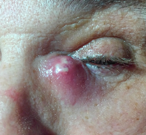
Acute dacryocystitis
Towards less invasive surgeries
In this study, an endoscopic approach was chosen for its high-definition visualization, precise navigation through distorted anatomy, and minimally invasive nature. Achieving the best results with this technique requires a thorough understanding of the anatomy. When combined with VR-assisted surgical planning, it allows for highly accurate and predictable outcomes.
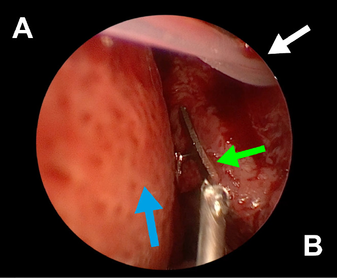
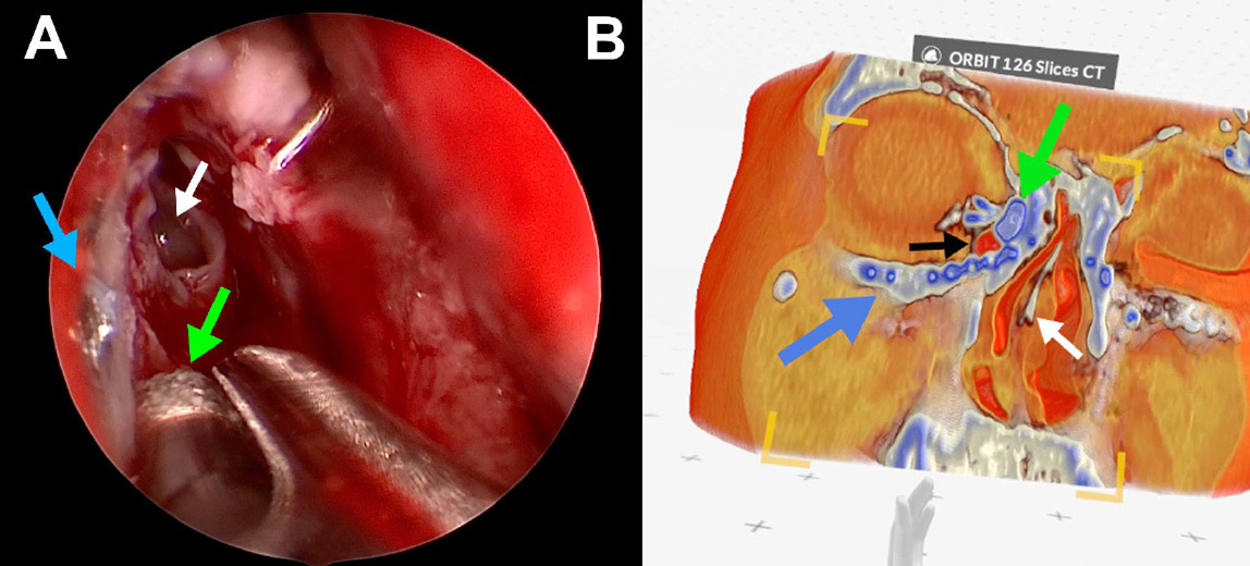
Endoscopic view of the left nasal cavity: white arrow — a silicone stent coming out from ostium, green arrow — orbital mesh, blue arrow — septal deviation (A — septum, B — lateral wall of the nasal cavity)
VR in surgical planning
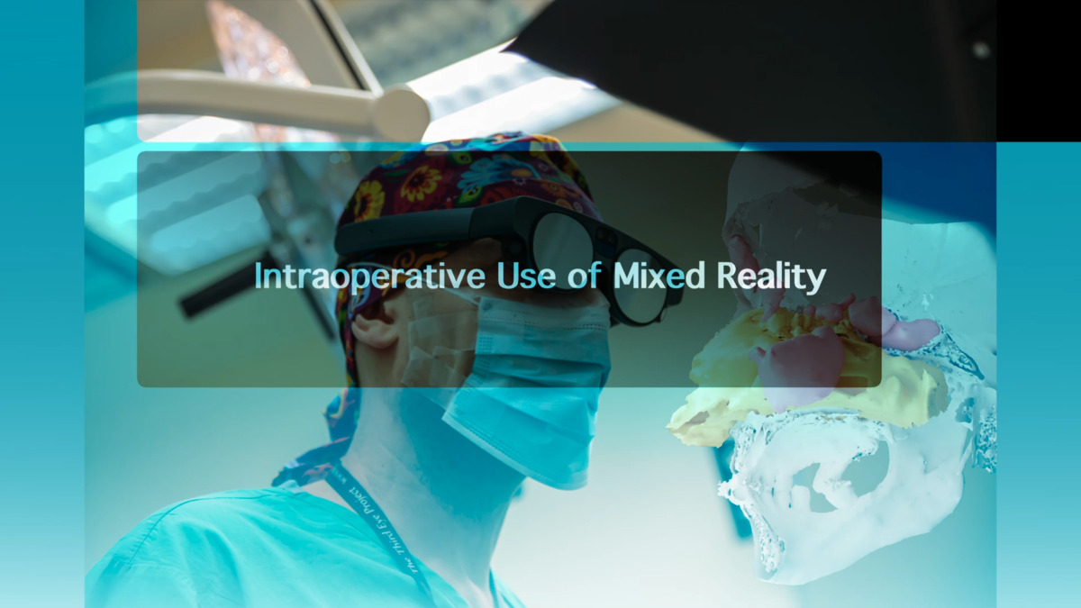
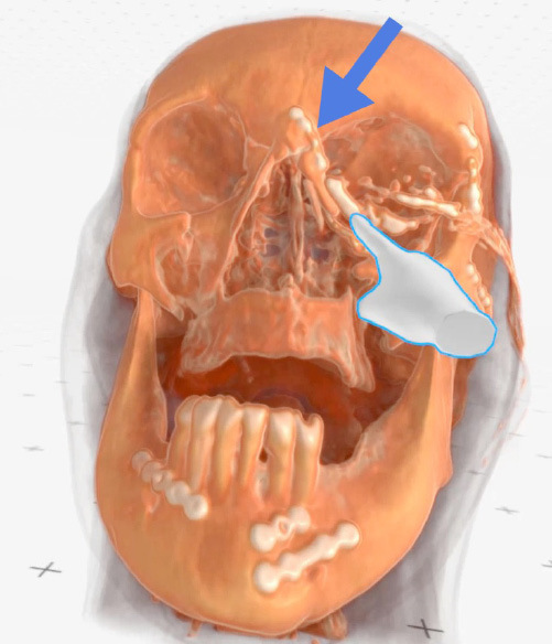
Using Medical Imaging XR, the surgical team generated detailed 3D reconstructions and examined them in an immersive VR environment. This allowed surgeons to examine the altered anatomy in 3D, manipulate structures freely, and gain an intuitive understanding of trauma-related distortions.
As a result, they were better able to anticipate potential challenges before surgery and refine their operative strategy with greater confidence. VR effectively bridged the gap between imaging and the surgical field, turning complex structures into a clear, navigable space.
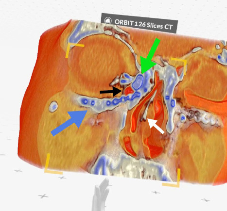
Case 2 (right side): green arrow — lacrimal sac, black arrow — air bubble inside the lacrimal sac, blue arrow — metal plate, white arrow — septal deviation (Medical Imaging XR reconstruction)
Safe and effective surgery
Over the observation period, the reconstructed tear drainage pathways remained functional, with no cases of recurrent tearing. The VR-supported workflow was safe, introduced no complications, and gave the surgical team a clearer, more predictable operative experience.
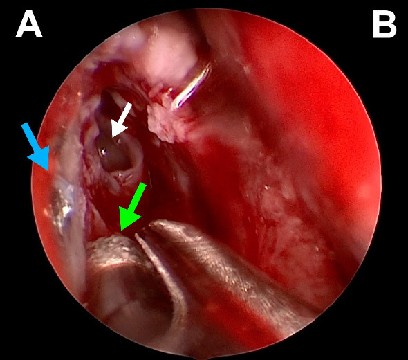
Endoscopic view of the right nasal cavity: green arrow — diamond burr, white arrow — opened agger nasi, blue arrow — tip of a metal bone screw (A — lateral wall of the nasal cavity, B — septum)
The future of surgical planning
This publication is among the first to integrate virtual reality directly into EnDCR planning, emphasizing its value in cases where trauma has significantly altered anatomy. The authors suggest that wider adoption could be supported by better technical documentation, standardized visual and surgical workflows, and ongoing improvements in software usability and design.
Check this out with Medicalholodeck
Medical Imaging XR enables clinicians to visualize, assess, and collaborate within an immersive, spatial environment. It allows real-time interaction with patient-specific 3D models, enhancing anatomical understanding, improving diagnostic accuracy, and supporting more precise surgical planning.
Medicalholodeck integrates with secure hospital systems, offering PACS access, HIPAA-compliant data handling, and full patient security. It works on VR headsets, PCs, iPads, and iPhones for flexible use in hospitals, classrooms, and training centers.
Specialized features for surgical planning are exclusive to Medical Imaging XR PRO.
Currently, Medicalholodeck is available only for educational use. The platform is undergoing FDA and CE certification, and we expect Medical Imaging XR PRO to be available soon in the U.S. and EU markets.
For more information, contact info@medicalholodeck.com February 2026