Download
Free fully functional 14-day trial included
Medicalholodeck AI supports healthcare, education, and patient care with advanced segmentation and immersive 3D visualization.
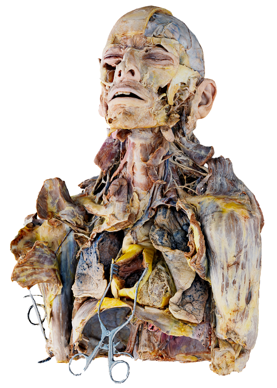
Educators and students gain interactive, anatomy-based learning with segmented 3D models from real patient data.
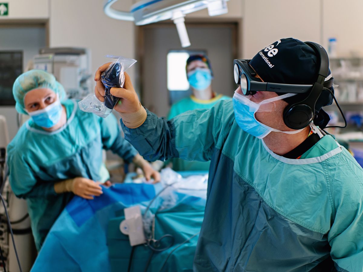
Surgeons use Medicalholodeck AI for precise planning, exploring segmented anatomy in 3D to better understand and prepare for complex procedures.*
Radiologists accelerate image interpretation with automated segmentation, gaining fast, structure-specific results that reduce manual effort and improve efficiency.*
Hospitals use 3D models to improve collaboration, support treatment decisions, and streamline daily workflows.*
Patients gain clearer insight into their diagnosis and treatment through 3D visualizations, improving consent and building trust with care teams.*
Generate segmented 3D models from CT or MRI scans in seconds. The AI detects anatomical structures automatically, saving time and reducing manual work.
Segment a wide range of organs and anatomical structures. Enable precise visualization, targeted analysis, and full-body exploration.
Access a growing library of AI models tailored to specific body regions and imaging types – including chest CT and whole-body scans. Choose the model that fits your clinical or educational use case.
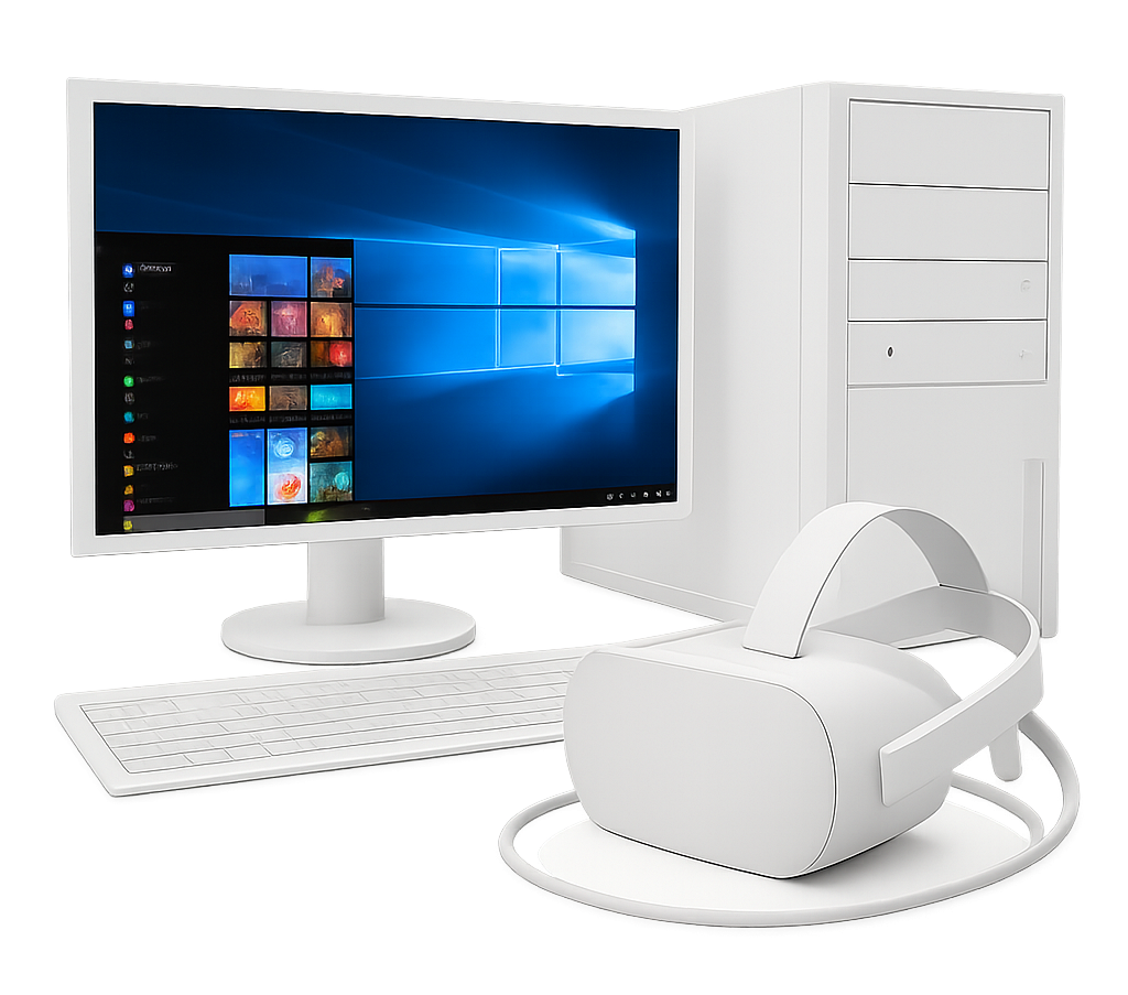
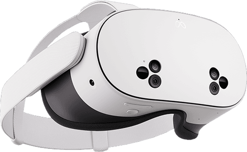
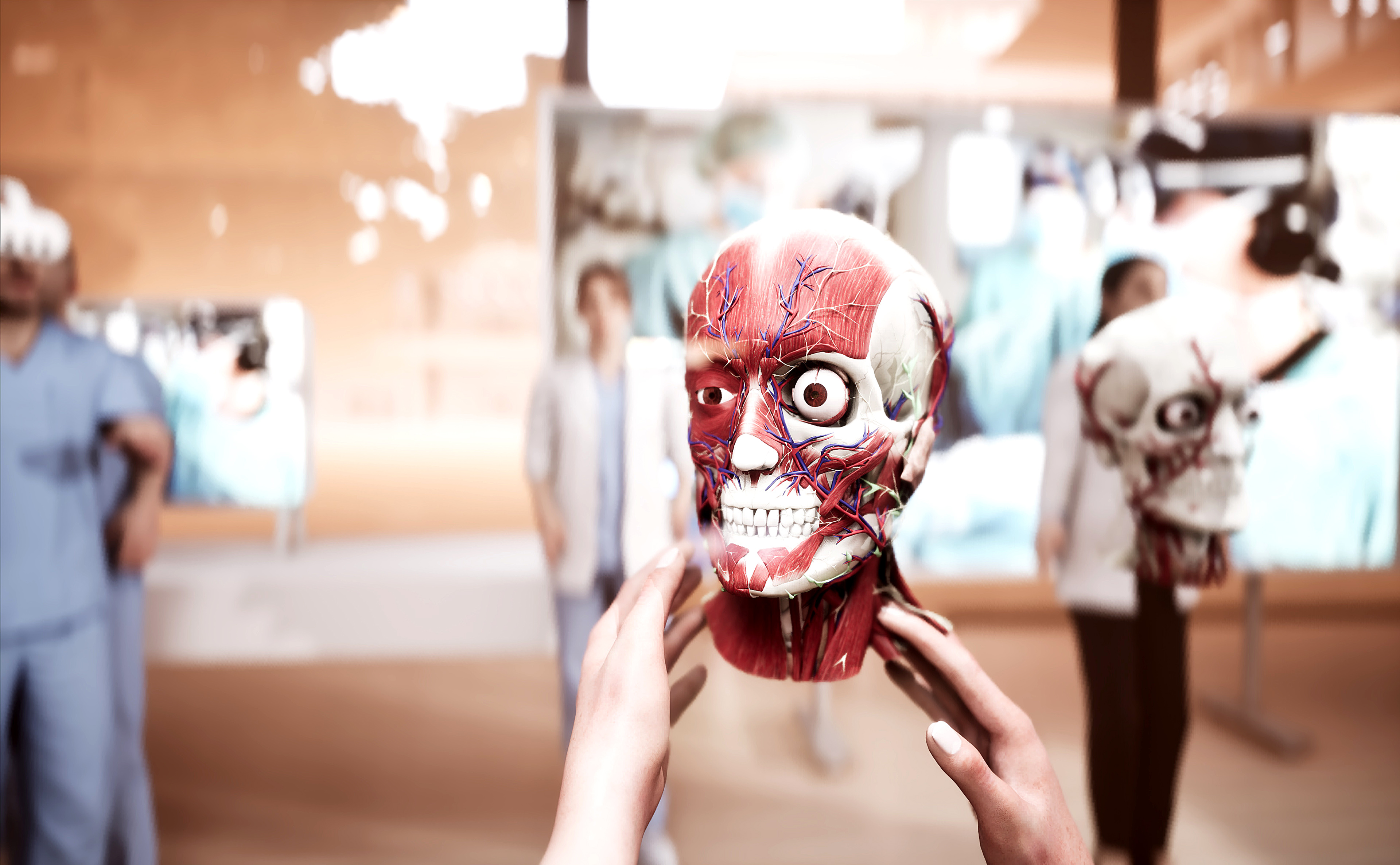
Explore segmented anatomy in immersive 3D using VR headsets, PCs, or mobile devices. Medicalholodeck offers intuitive, spatial interaction with patient-specific data to support deeper understanding and clearer communication.
Plan complex procedures with patient-specific, anatomically accurate 3D models that enhance spatial understanding and support better outcomes.*
Integrate real-patient anatomy into classrooms, labs, and simulations. Ideal for teaching anatomy, radiology, and surgical workflows in immersive environments.
Use clear, interactive 3D models to help patients understand their condition and treatment options, improving communication, consent, and trust.*
AI segmentation automatically detects and separates anatomical structures like organs, bones, and tissues from CT or MRI scans. Instead of drawing each region by hand, AI models complete the task in seconds by recognizing patterns from thousands of expert-labeled images.
Built using deep learning – typically convolutional neural networks (CNNs) – these models are trained on large datasets of anonymized DICOM scans. Once trained, they can accurately segment new data, structure by structure.
This speeds up radiological analysis, surgical planning, and 3D model creation, while improving accuracy and team communication. In VR or AR, it enables clear, spatial understanding of complex anatomy.
Medicalholodeck AI turns slow, manual segmentation into a fast, precise, and immersive process – helping professionals work more efficiently and make informed decisions.
Medicalholodeck supports a growing range of high-performance segmentation models, developed by leading institutions and AI frameworks, to deliver accurate and efficient results across diverse medical imaging needs.
The following models are currently supported and continuously updated:
Robust detection of 117 organs and structures in CT images (1.5mm resolution). Model created by the department of Research and Analysis at University Hospital Basel and made publicly available as part of TotalSegmentator.
Fast & robust detection of 117 organs and structures in CT images (3m resolution). Model created by the department of Research and Analysis at University Hospital Basel and made publicly available as part of TotalSegmentator.
Robust detection of 56 organs and structures in MR images (1.5mm resolution). Model created by the department of Research and Analysis at University Hospital Basel and made publicly available as part of TotalSegmentator.
Robust detection of lung vessels, trachea and bronchia. Model created by the department of Research and Analysis at University Hospital Basel and made publicly available as part of TotalSegmentator.
Robust detection of the body, trunc, skin and extremities. Model created by the department of Research and Analysis at University Hospital Basel and made publicly available as part of TotalSegmentator.
Detection of head glands cavities. Model created by the department of Research and Analysis at University Hospital Basel and made publicly available as part of TotalSegmentator.
Detection of head muscles. Model created by the department of Research and Analysis at University Hospital Basel and made publicly available as part of TotalSegmentator.
Detection of neck bones and vessels. Model created by the department of Research and Analysis at University Hospital Basel and made publicly available as part of TotalSegmentator.
Detection of neck muscles. Model created by the department of Research and Analysis at University Hospital Basel and made publicly available as part of TotalSegmentator.
Detection of coronary arteries. These models are not trained on the full totalsegmentator dataset but on some small other datasets. Therefore, expect them to work less robustly. Model created by the department of Research and Analysis at University Hospital Basel and made publicly available as part of TotalSegmentator.
Detection of intracerebral hemorrhage. These models are not trained on the full totalsegmentator dataset but on some small other datasets. Therefore, expect them to work less robustly. Model created by the department of Research and Analysis at University Hospital Basel and made publicly available as part of TotalSegmentator.
Detection of pleural effusion and pericardial effusion. These models are not trained on the full totalsegmentator dataset but on some small other datasets. Therefore, expect them to work less robustly. Model created by the department of Research and Analysis at University Hospital Basel and made publicly available as part of TotalSegmentator.
Detection of hip implants. These models are not trained on the full totalsegmentator dataset but on some small other datasets. Therefore, expect them to work less robustly. Model created by the department of Research and Analysis at University Hospital Basel and made publicly available as part of TotalSegmentator.
Detection of liver vessels and tumor. These models are not trained on the full totalsegmentator dataset but on some small other datasets. Therefore, expect them to work less robustly. Model created by the department of Research and Analysis at University Hospital Basel and made publicly available as part of TotalSegmentator.
Detection of oculomotor muscles. These models are not trained on the full totalsegmentator dataset but on some small other datasets. Therefore, expect them to work less robustly. Model created by the department of Research and Analysis at University Hospital Basel and made publicly available as part of TotalSegmentator.
Robust segmentation of 104 anatomical structures in CT images. Model created by MONAI team and made publicly available as part of MONAI Model Zoo.
Segmentation with 133 structures from T1W MRI image. Model created by Vanderbilt University & MONAI team and made publicly available as part of MONAI Model Zoo.
Multi-organ segmentation of 13 structures from CT image. Model created by MONAI team and made publicly available as part of MONAI Model Zoo.
Segmentation of the pancreas and pancreatic tumor from CT image. Model created by MONAI team and made publicly available as part of MONAI Model Zoo.
Segmentation of the prostate from MRI images. Model created by Keno Bressem and made publicly available as part of MONAI Model Zoo.
Segmentation of the spleen from CT images. Model created by MONAI team and made publicly available as part of MONAI Model Zoo.
Segmentation of the spleen from CT images using DeepEdit. Model created by MONAI team and made publicly available as part of MONAI Model Zoo
Detection of trunc and extremities. Model created by the department of Research and Analysis at University Hospital Basel and made publicly available as part of TotalSegmentator.
Detection of vertebrae. Model created by the department of Research and Analysis at University Hospital Basel and made publicly available as part of TotalSegmentator.
Detection of 8 liver segments. Model created by the department of Research and Analysis at University Hospital Basel and made publicly available as part of TotalSegmentator.
Detection of kidney cysts. Model created by the department of Research and Analysis at University Hospital Basel and made publicly available as part of TotalSegmentator.
Detection of breasts. Model created by the department of Research and Analysis at University Hospital Basel and made publicly available as part of TotalSegmentator.
Detection of lung and lung nodules. Model provided by BLUEMIND AI: Fitzjalen R., Aladin M., Nanyan G. and made publicly available as part of TotalSegmentator.
Medicalholodeck offers intuitive tools for fast, efficient, and collaborative work with segmented medical data – whether you're planning surgery, teaching, or reviewing clinical cases.*
Quickly toggle the visibility of individual organs or regions to isolate specific anatomy and focus your analysis.
Save time and ensure consistency with Smart Presets by storing AI and visualization settings.
Perform precise 3D measurements directly within the model for accurate and detailed analysis.
Display and compare multiple datasets side by side to assess changes and track outcomes over time.
Collaborate in real time with colleagues, students, or remote teams. Share sessions to explore and discuss cases together in a virtual environment.
Capture sessions with the integrated RXR feature for easy playback. Save your work, export settings, and securely share cases locally or across institutions.
Medicalholodeck supports custom model integration, allowing you to use institution-trained or third-party AI engines. Contact us to discuss your setup.
Integration options
Use on cloud
Run segmentation securely on the Medicalholodeck cloud – no installation needed, always up to date, and accessible across devices.
Use local
Keep all data in-house with local segmentation on high-performance PCs or servers – no internet required.
Enterprise integration options
Integrate with hospital systems, PACS, or custom AI models. API access and on-premise deployment available.
For setup and deployment support, contact us at support@medicalholodeck.com
Speed
AI segmentation typically completes in under 60 seconds, providing near-instant access to high-quality 3D models and streamlining clinical and educational workflows.
Accuracy
Medicalholodeck AI models are trained on large, expert-annotated datasets to ensure consistent, high-precision results across standard imaging scenarios.
Compliance
All processing is designed with data security in mind and adheres to strict privacy regulations – fully aligned with GDPR and HIPAA requirements for safe clinical use.