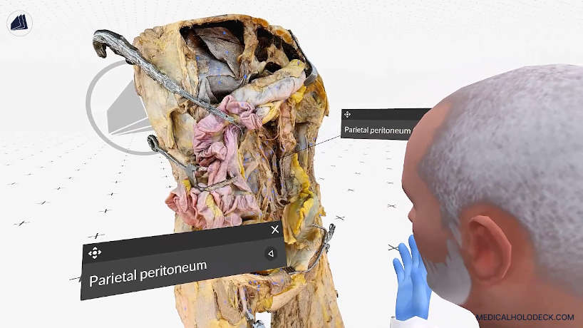
Abdomen and pelvis
Explore the anatomical layers of the abdomen and pelvis, including visceral and retroperitoneal structures.
Medicalholodeck Academy
Review cranial and thoracic anatomy through a guided exploration of layered structures.
This video is a preview. Download the RXR recording for interactive playback in Medicalholodeck.
Lesson overview
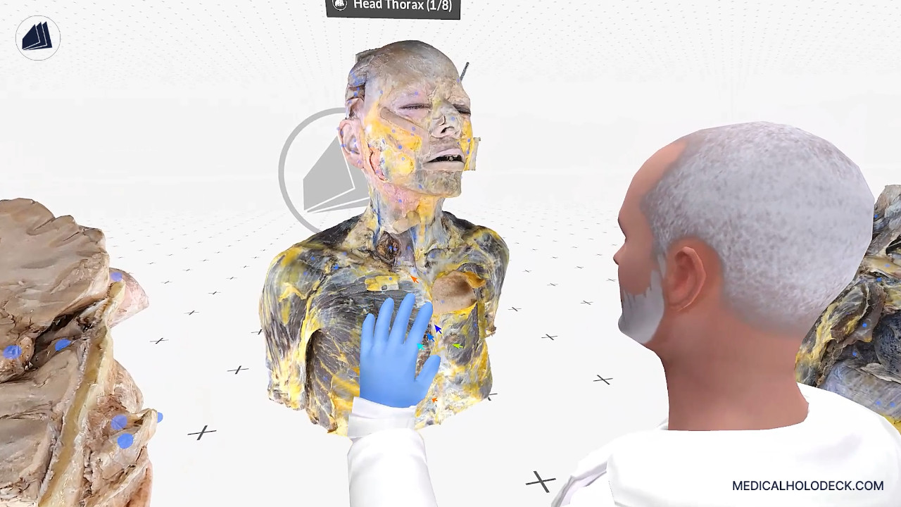
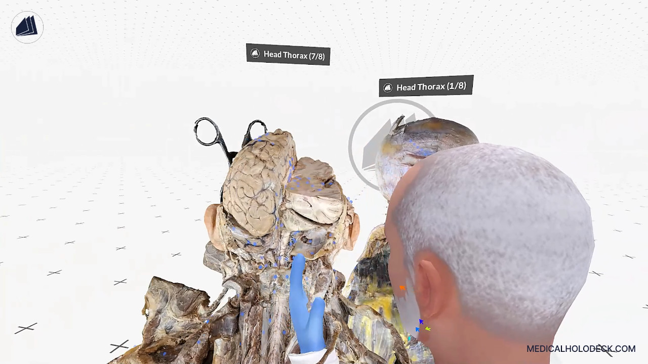
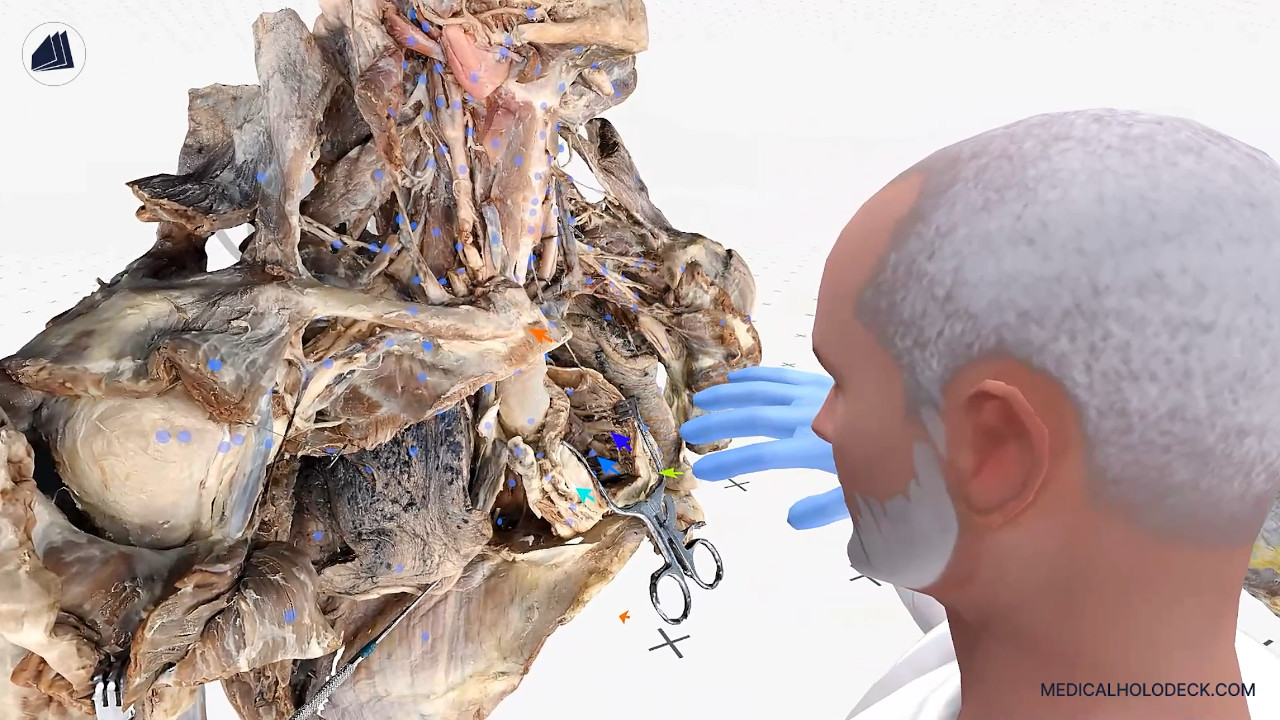
Explore the chest wall, thoracic cavity, face, and neck, then examine deeper structures including the brain and cranial nerves. Follow the anatomical continuity between the thorax and neck.
In this lesson, Evan guides you through the head and thorax model, exploring key anatomical regions such as the chest wall, thoracic cavity, face, neck, and deeper structures including the brain and cranial nerves. He highlights the axillary region with the brachial plexus, shoulder, back, and spinal cord on deeper dissections.
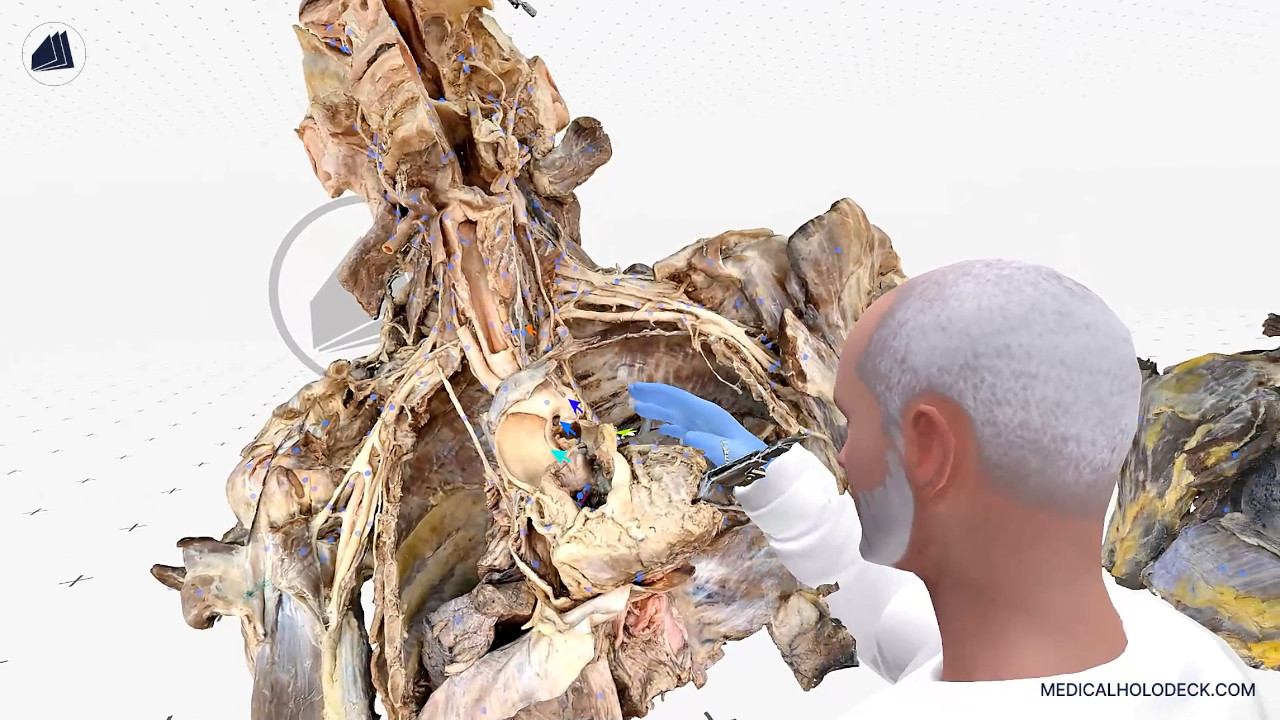
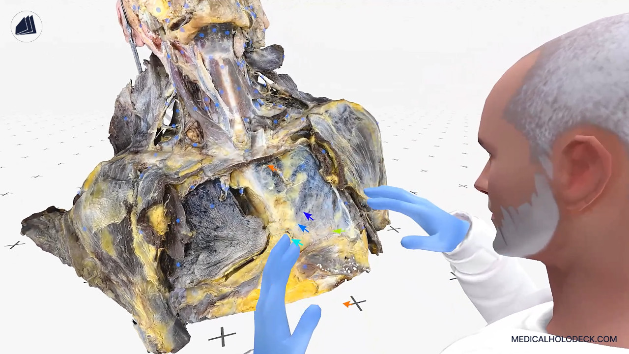
Evan demonstrates how removing the chest wall reveals the lungs and parietal pleura, while further layers expose the heart with visible coronary circulation, papillary muscles, chordae tendineae, valves, and the parietal pericardium. Important nerves like the phrenic and vagus nerves, and major vessels such as the brachiocephalic artery, are also shown.
Deeper views include the ascending aorta, interior of aortic arch, heart chambers, and lung root structures such as bronchi and pulmonary vessels. The deepest layer reveals the posterior mediastinum with intercostal nerve vasculature, sympathetic chain, azygos vein, thoracic aorta, and splanchnic nerve. Finally, Evan illustrates the continuity between thoracic structures and the vessels extending into the neck.
Inside the lesson
Interactive learning
Download the RXR file and open it in Medicalholodeck. Replay the explanation, pause to examine a structure, and explore the anatomy from different viewpoints.
New to Medicalholodeck? Download a trial to get started.
Continue learning
Explore the anatomical layers of the abdomen and pelvis, including visceral and retroperitoneal structures.
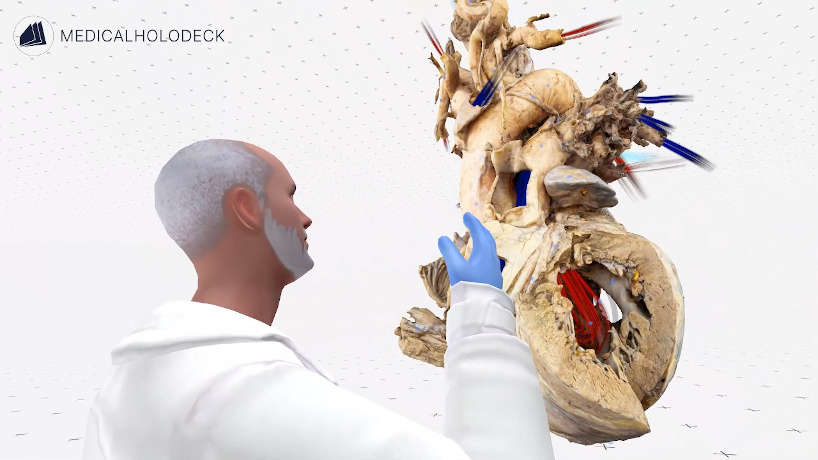
Follow blood flow through the heart and examine the structures involved in circulation.
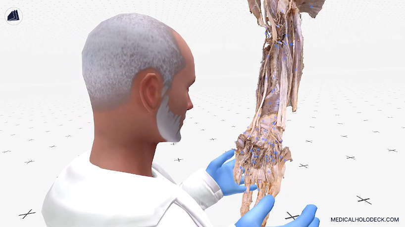
Trace muscles, nerves, and vessels from the upper arm to the hand.