Radiology provides detailed images of internal anatomy, making it fundamental for diagnosing, monitoring, and treating a wide range of medical conditions. With spatial imaging, conventional 2D imaging from CT, MRI, CBCT, ultrasound, and other DICOM-formatted scans is transformed into interactive 3D digital twins. Clinicians can explore patient-specific anatomy in an immersive environment, gaining deeper insights into complex structures.
Important notice regarding surgical planning and professional clinical use
Specialized functionalities for surgical planning and pre-operative professional applications are exclusive to Medical Imaging XR PRO FDA. This version is not yet available. Information about the release date will be published here soon.
Medicalholodeck is currently undergoing the required FDA (U.S. Food and Drug Administration) and CE (Conformité Européenne) certification processes. Our team is working diligently to ensure full compliance with all regulatory standards, and we expect Medical Imaging XR PRO to be available in both the United States and the European Union soon.
For updates on product releases, regulatory progress, and availability, or for any related inquiries, please contact info@medicalholodeck.com.
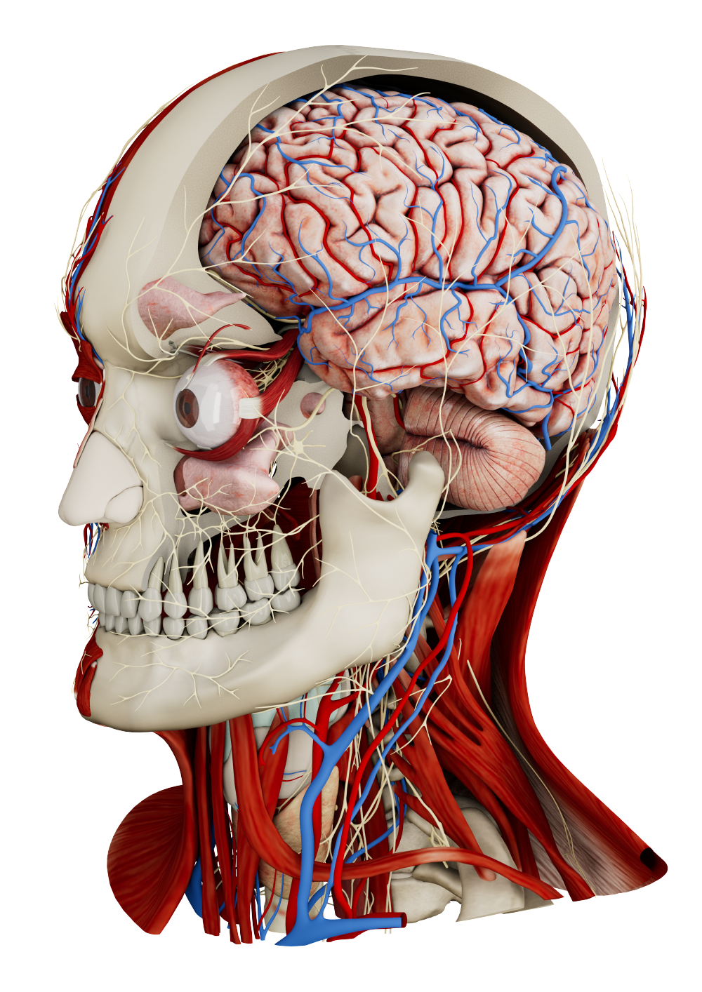
From 2D to 3D: Get the full picture
Radiology creates and interprets images of the inside of the body to guide clinical decision-making, surgical planning, and medical education. Virtual reality enhances this process by converting 2D scans into 3D reconstructions that can be explored in a spatial environment. Users can step inside the patient’s anatomy with true depth and spatial context, rotating, slicing, and zooming to highlight bones, vessels, nerves, and pathological areas.
With integrated AI-powered segmentation, critical structures are automatically identified and separated, reducing preparation time and allowing radiologists to focus on clinical interpretation.
Better imaging, better results
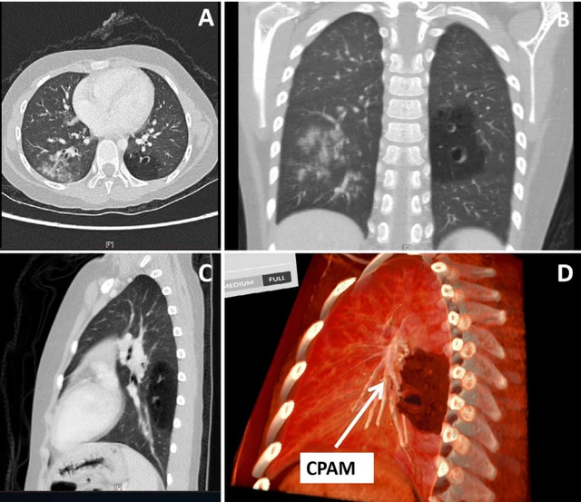
Yang W, Xu Y, Wang Z, Ye M, Chen R, Da M, Qi J (2025) Virtual reality-assisted preoperative planning for pediatric thoracoscopic segmentectomy: a retrospective study. BMC Pediatrics.
3D visualization of radiological data enables precise localization of abnormalities and structures, improving diagnostic accuracy and surgical planning. Interactive 3D models streamline access planning, allowing surgeons and radiologists to explore optimal routes and reduce risks. Real-time multi-user sessions support collaborative case discussions, letting teams share insights and agree on the best approach – whether in the same hospital or across continents.
Read more: https://doi.org/10.1186/s12887-025-06259-3
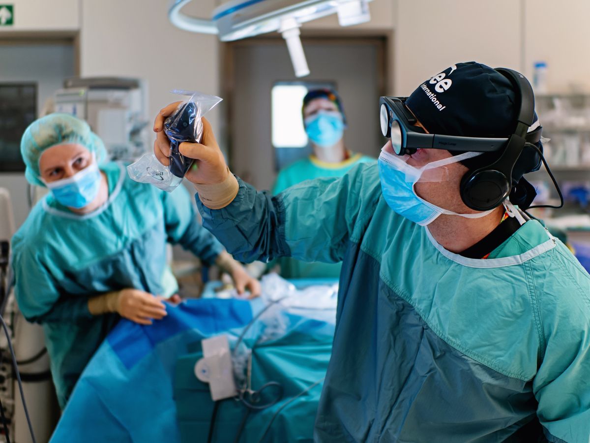
Medical visual communication
Radiologists, surgeons, assistants, and other medical professionals can discuss, plan, and rehearse surgical procedures together using shared 3D visualizations. This collaborative approach improves understanding, coordination, and precision throughout the surgical workflow.
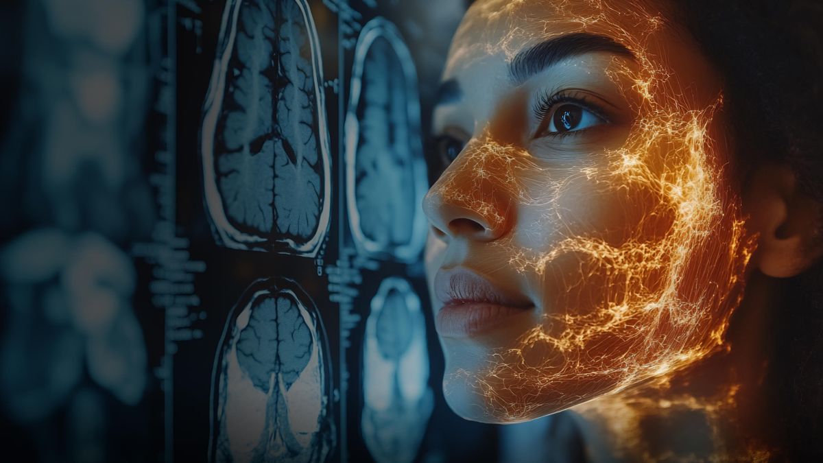
Diagnostics
3D imaging enhances diagnostic accuracy by providing detailed volumetric views that go beyond traditional 2D scans. Radiologists can visualize complex pathologies from multiple angles, detect subtle abnormalities, and differentiate overlapping structures with greater precision. Advanced rendering techniques and AI integration further support early detection, measurement, and characterization of diseases.
Surgical planning
The 3D interactive models accurately replicate real-world anatomical structures, enabling surgeons to simulate procedures, explore different surgical approaches, and anticipate potential complications before entering the operating room.
Read more about surgical planning
Spatial telemedicine and collaboration
XR and AI-powered 3D imaging enable surgeons and radiologists to work together globally in real time, sharing interactive visualizations and collaboratively annotating scans beyond geographical boundaries.
Patient information
Patient information is about helping people understand their medical situation. With 3D imaging, doctors can turn complex scans into clear, easy-to-follow visuals of a patient’s body. This makes it easier to see what’s happening, understand the diagnosis, and explore treatment options. It also reduces anxiety by giving patients a sense of control through a clearer understanding of their condition.
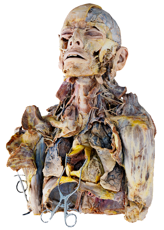
Medical training
Medical educators and trainees gain hands-on experience with real radiological datasets in immersive 3D environments. By exploring anatomy interactively, they deepen their understanding of complex structures, practice clinical procedures, and develop essential skills for real-world medical practice.
How to get started
Medicalholodeck integrates with secure hospital systems, providing PACS access, HIPAA-compliant data handling, and full patient data security. It runs on stereoscopic 3D displays, VR headsets, mobile devices, and standard Windows systems, enabling flexible use in hospitals, classrooms, and training centers.
Specialized features for surgical planning are exclusive to Medical Imaging XR PRO. Currently, Medicalholodeck is available only for educational use. The platform is undergoing FDA and CE certification, and we expect Medical Imaging XR PRO to be available soon in the U.S. and EU markets.
For updates, regulatory news, availability, or questions contact info@medicalholodeck.com.