MEDICALHOLODECK®
The human body is not flat. Yet medical imaging and anatomy are still primarily interpreted in two dimensions, creating a gap between data and understanding.
Medicalholodeck closes this gap by converting CT, MRI, and DICOM data into interactive 3D volumes, enabling direct spatial analysis of anatomy and pathology. The result is improved precision, faster interpretation, clearer team communication, and better-trained medical professionals.
Apps
Medicalholodeck brings together a suite of apps that power spatial medicine across clinical practice, education, and collaboration.
Use cases
Medicalholodeck supports professional and educational use. It is used for anatomy teaching, simulation, and training with real medical data in interactive 3D. Users explore structures in context and improve spatial understanding.
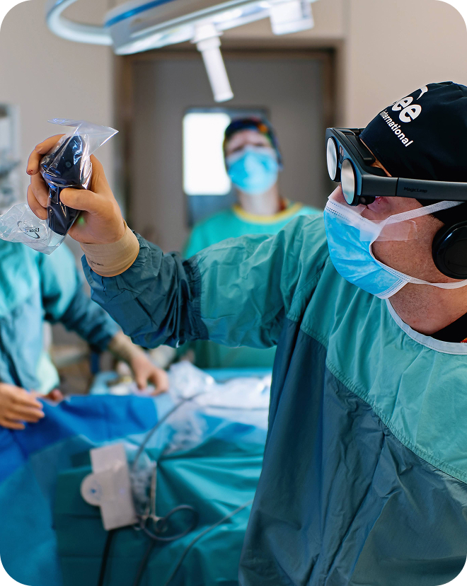
In clinical environments, it supports case discussions, tumor boards, and interdisciplinary collaboration using CT and MRI data. Teams review the same dataset in real time. This improves clarity, reduces misinterpretation, and speeds up alignment.
It also enables creation of patient-specific digital twins for training and preparation. Clinicians analyze cases, test approaches, and prepare procedures. It improves patient communication and supports remote collaboration and session recording.*
Advantages for professionals
Work with CT and MRI data in true 3D to improve how anatomy and pathology are understood, analyzed, and communicated. Interactive exploration, AI-supported digital twins, and immersive training enable faster and more accurate decision-making.
Immersive imaging: Work with medical imaging in true 3D and gain deeper clinical insight. Move beyond flat screens and interact with patient data in immersive environments.
Impact: Faster comprehension reduces analysis time.
Interactive data: Explore CT and MRI as interactive 3D volumes. View anatomy and pathology from any angle, perform precise measurements, enabling more accurate analysis.
Impact: Improved accuracy reduces treatment costs.
Advanced procedure training: Train procedures with patient-specific digital twins. Simulate interventions in advance to identify optimal approaches.
Impact: Better preparation shortens operating room time.
Reduced surgical risk: Reduce procedure time and complications through better preparation. Precise training enables more efficient workflows and improved outcomes.
Impact: Fewer complications reduce post-operative costs.
Clear clinical communication: Communicate complex information with clarity for both patients and care teams.
Impact: Clear communication reduces delays and misunderstandings.
Seamless team collaboration: Collaborate across teams and locations in shared immersive sessions. Discuss cases with precise spatial understanding in real time.
Impact: Faster alignment between specialists speeds up treatment.
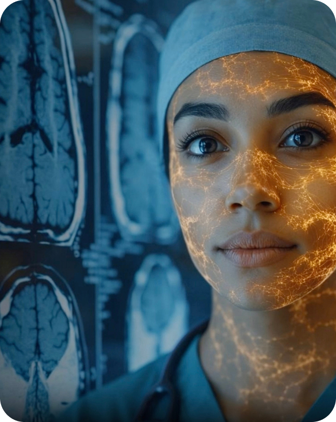
Advantages for education
Learn anatomy and imaging in true 3D with real data and AI. Students explore, practice safely, and learn interactively. The result is faster learning, better understanding, and lower infrastructure costs.
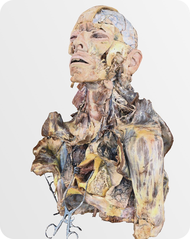
3D anatomy learning: Explore anatomy models and dissections in true 3D. Understand structures in context.
Impact: Faster comprehension shortens learning cycles and improves efficiency.
Digital twins and AI: Use CT and MRI datasets to study real cases, automatically segmented for rapid analysis.
Impact: Learning from everyday cases accelerates learning curves.
Immersive education: Teach, train, and learn with real medical data and anatomy in fully interactive 3D.
Impact: Reduces reliance on physical labs and materials, lowering infrastructure costs.
Spatial medical understanding: Develop a deeper understanding of spatial relationships in anatomy and pathology.
Impact: Better-trained professionals make fewer errors.
Interactive teaching: Instructors guide students through anatomy and cases in real time, enabling engaging and dynamic lessons.
Impact: More effective teaching reduces repetition and time.
Safe trainings: Allow students to make mistakes and learn without risk to real patients.
Impact: Reduces early-stage training errors and improves readiness.
Hardware
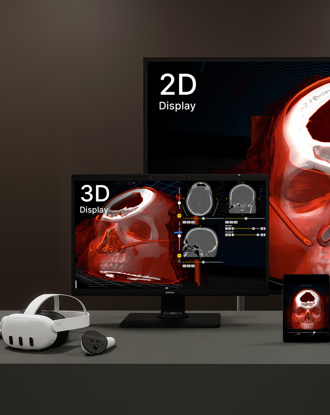
Medicalholodeck supports a wide range of hardware setups, from high-performance VR and PC systems to standalone VR headsets and mobile devices.
Choose between high-performance 3D setups and simpler options for education or mobility, based on your needs and budget. Advanced setups use a high-performance PC with VR headsets, 3D displays, and standard screens at the same time. This enables full-resolution imaging, AI segmentation, and synchronized use across devices.
Simpler setups include standalone VR and mobile devices, which are easier to deploy but less powerful. Remote rendering runs processing on a server and streams to devices, combining high quality with flexible access. See all hardware setup options here.
Solutions and plans
Medicalholodeck offers plans for students, schools, universities, hospitals, and physicians, designed for different experience levels – from basic learning to professional use.
Plans support education and clinical workflows, from anatomy learning to working with real patient data and cases. Start with what you need and scale over time for individuals, classrooms, or clinical teams.
Explore all features and plans or get in touch with our team for guidance.