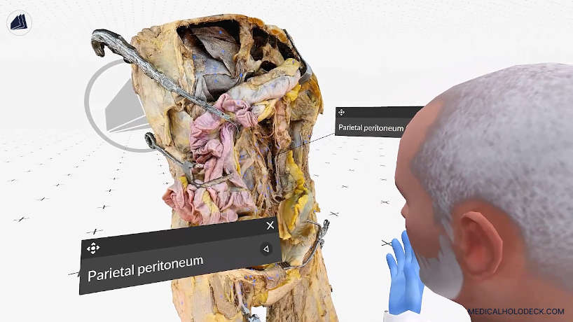
Abdomen and pelvis
Explore the anatomical layers of the abdomen and pelvis, including visceral and retroperitoneal structures.
Medicalholodeck Academy
Examine the nerves, arteries, and anatomical relationships of the hand.
This video is a preview. Download the RXR recording for interactive playback in Medicalholodeck.
Lesson overview
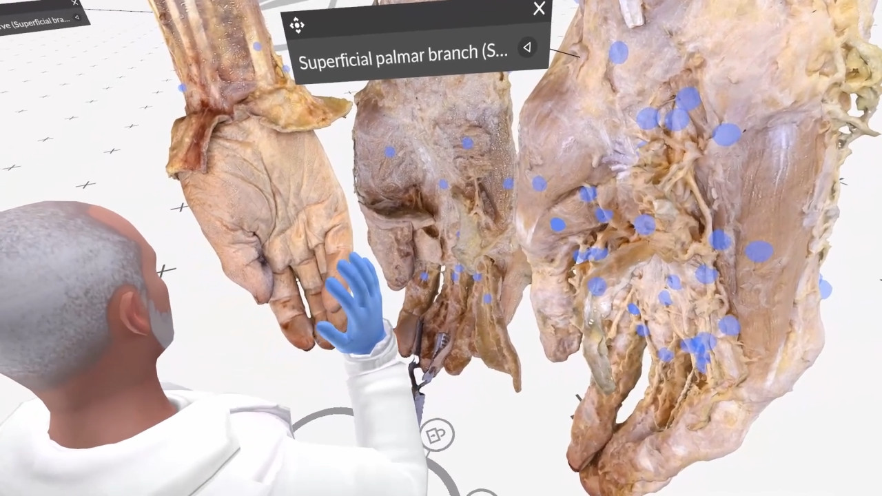
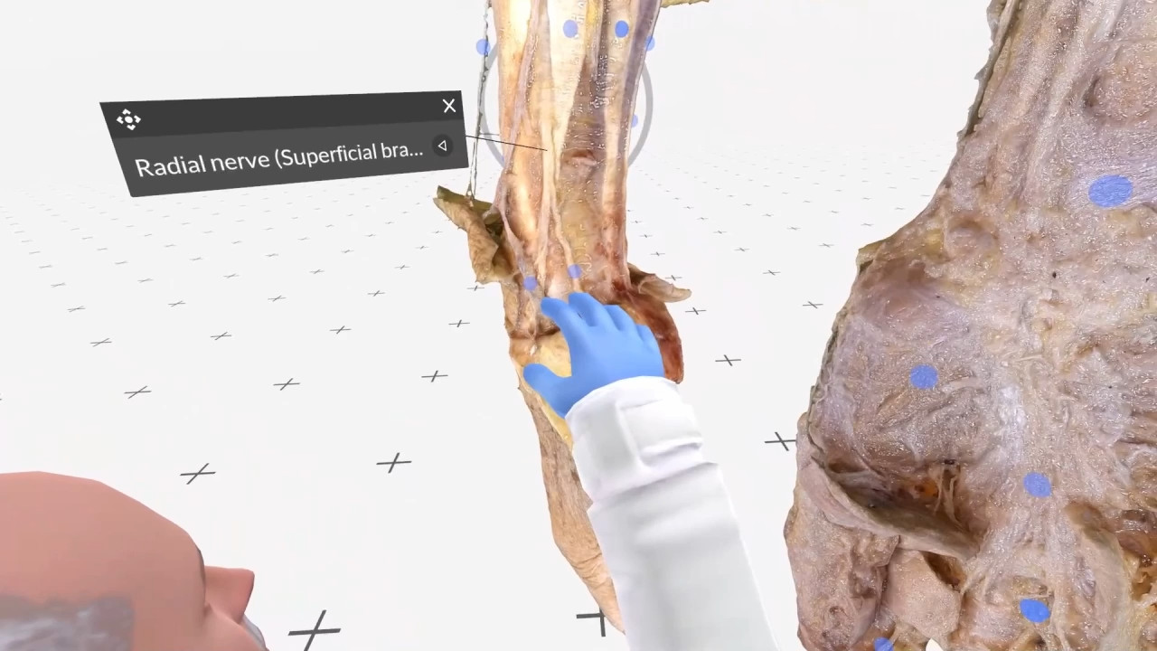
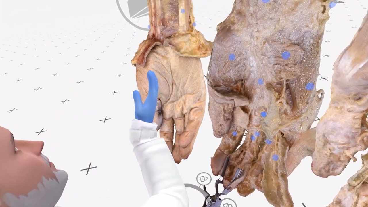
Review the anatomical layers of the hand, beginning with superficial nerves and veins. Examine deeper structures, including the palmar aponeurosis and flexor retinaculum.
In this VR lesson, Evan highlights important anatomical features encountered during initial hand dissections. Just beneath the skin, you can identify superficial structures such as the superficial branch of the radial nerve and the cephalic vein. Evan points out a subtle bump that may seem insignificant superficially but, upon deeper dissection, reveals itself as a branch of the radial artery, underscoring the importance of careful layering to avoid damaging vital vessels.
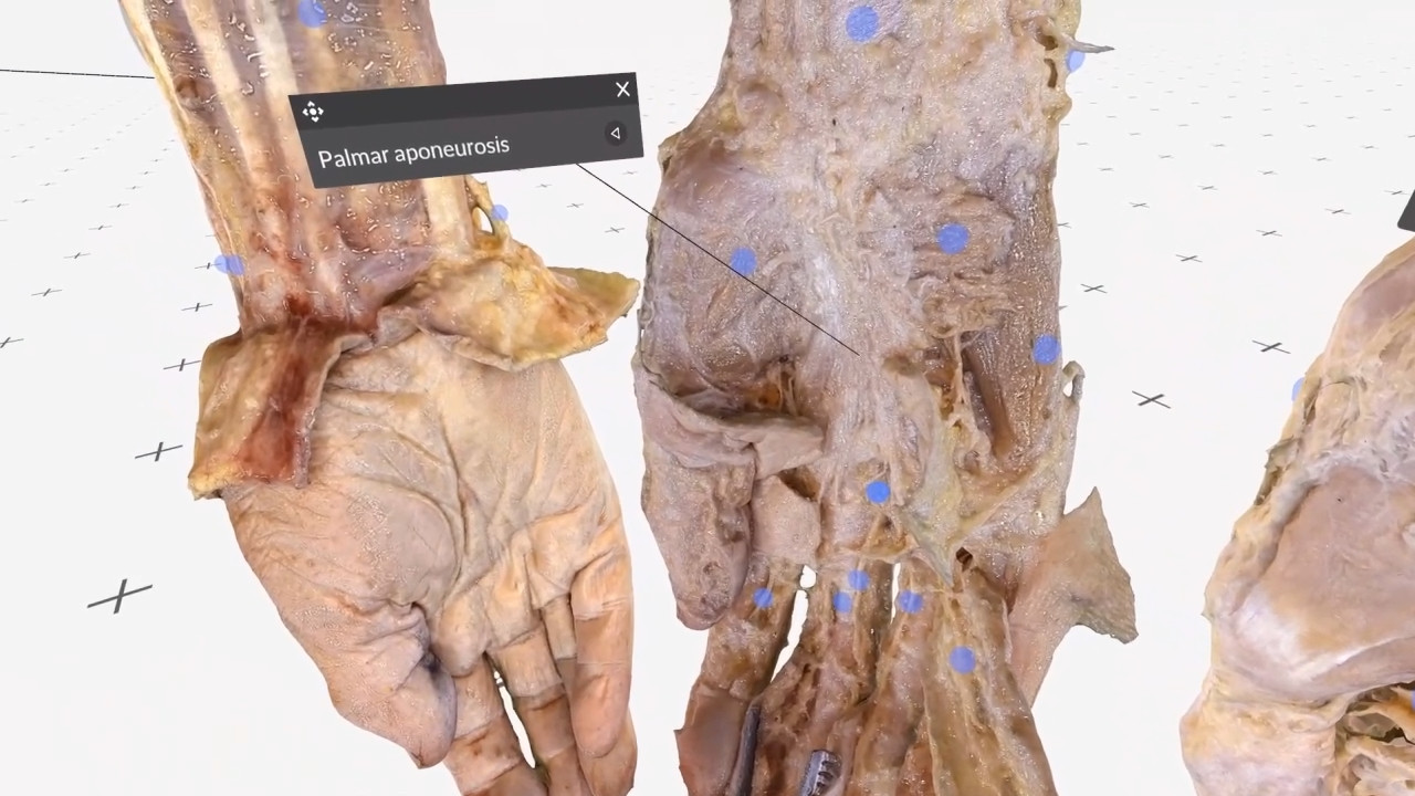
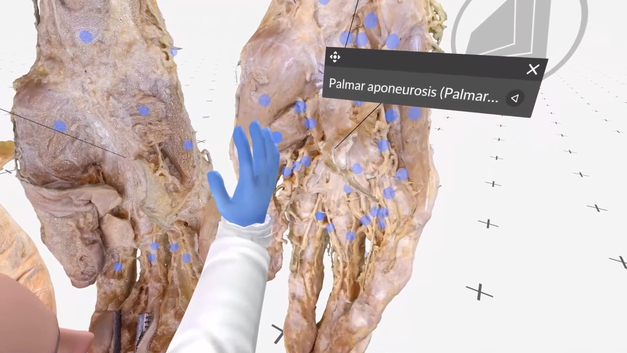
He then focuses on the palmar aponeurosis, a prominent superficial fascial structure of the palm. Beneath this lies the flexor retinaculum, visible after the aponeurosis is cut, serving as a landmark for further exploration. Evan emphasizes how many branches of nerves and arteries pass deep to these fascial layers, illustrating key considerations for safely navigating the complex anatomy of the hand.
Inside the lesson
Interactive learning
Download the RXR file and open it in Medicalholodeck. Replay the explanation, pause to examine a structure, and explore the anatomy from different viewpoints.
New to Medicalholodeck? Download a trial to get started.
Continue learning
Explore the anatomical layers of the abdomen and pelvis, including visceral and retroperitoneal structures.
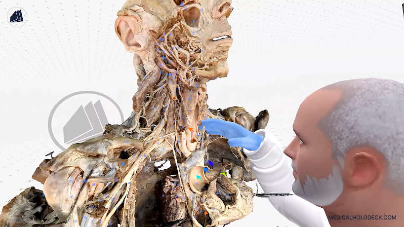
Review cranial and thoracic anatomy through a guided exploration of layered structures.
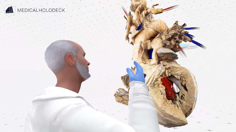
Follow blood flow through the heart and examine the structures involved in circulation.