Integrating Artificial Intelligence Into the Visualization and Modeling of Three-Dimensional Anatomy in Pediatric Surgical Patients. Journal of Pediatric Surgery, 2024; 59
https://doi.org/10.1016/j.jpedsurg.2024.07.014Manual and AI segmentation
Medical image segmentation aims to divide images into meaningful regions to isolate anatomical structures, detect abnormalities, and provide measurements for diagnosis, treatment planning, and disease monitoring. Algorithmic tools can accelerate this process, enabling 3D rendering or export within minutes. In this study, 3D Slicer was used to fine-tune the segments, although most 3D models were generated automatically using AI extensions.
AI segmentation tools
Applications for preoperative planning increasingly use AI algorithms to analyze medical images.
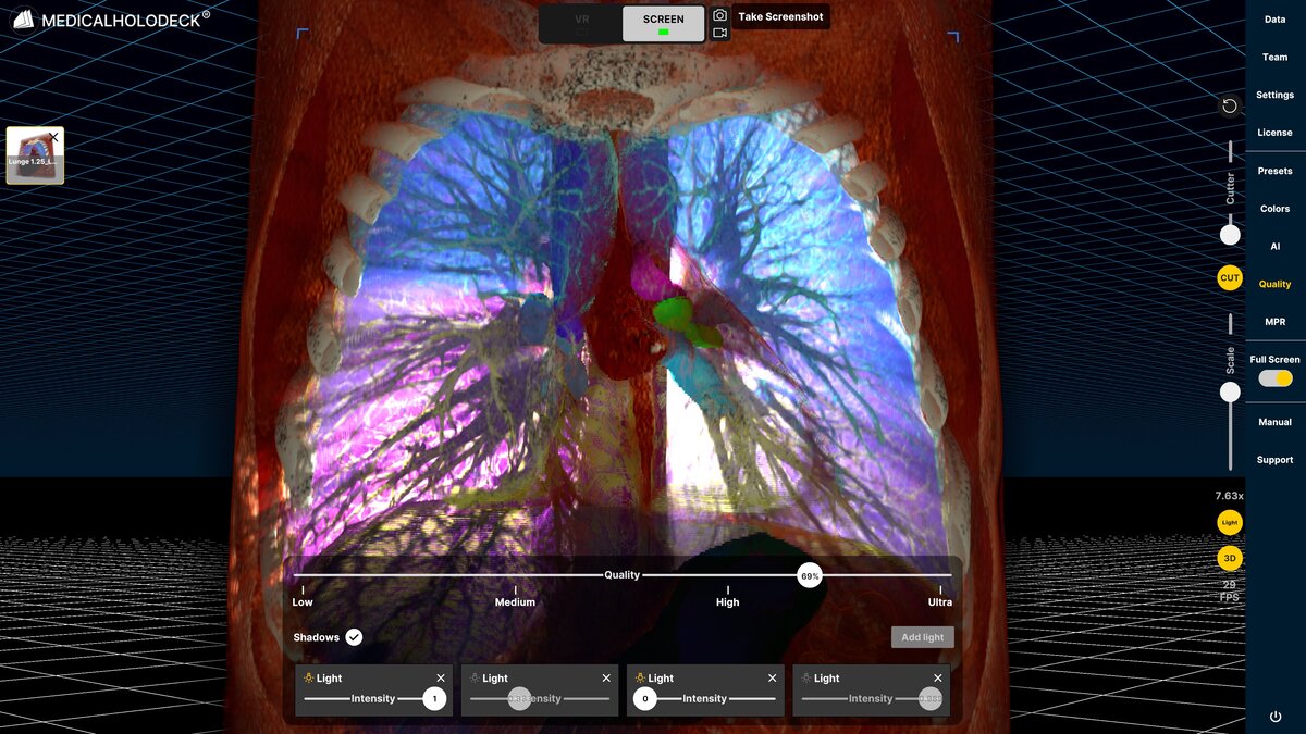
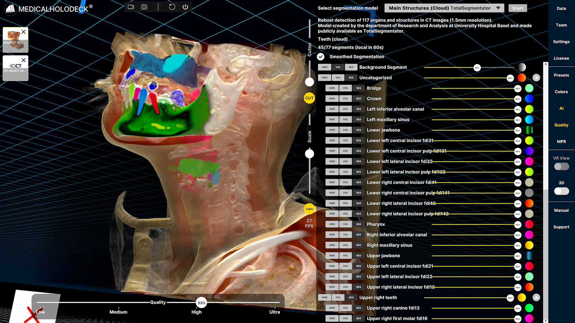
TotalSegmentator, trained on 1,204 CT series from University Hospital Basel, can segment 104 anatomical structures with high precision. It is integrated into 3D Slicer as well as commercial 3D/VR platforms like Medicalholodeck.
MONAI is an open-source deep learning framework for healthcare. Its 3D Slicer extension, MONAI Auto3DSeg, provides automated segmentation models for specific organs and enables researchers to train custom models for targeted pathologies.
Patient MRI and CT scans were de-identified and exported in DICOM format for testing AI extensions in 3D Slicer, using a convenience sample of pediatric surgical cases.
AI in pediatric 3D planning
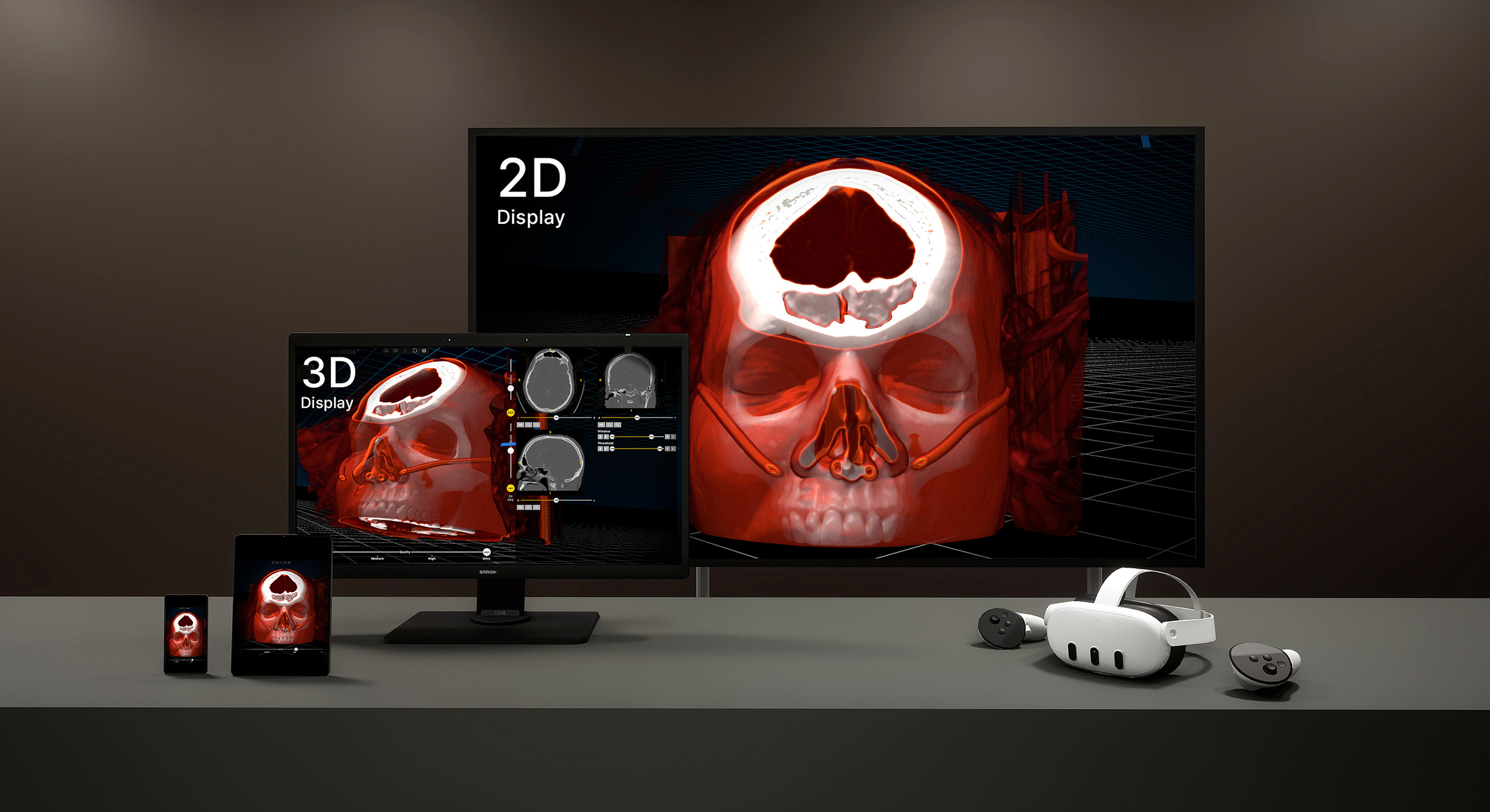
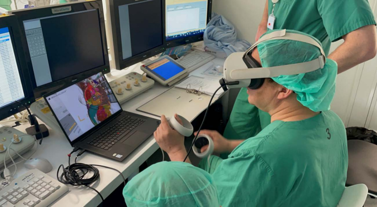
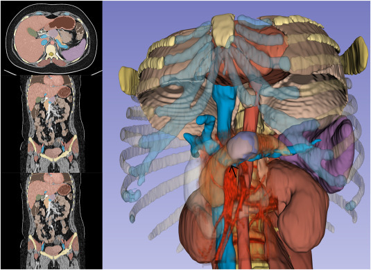
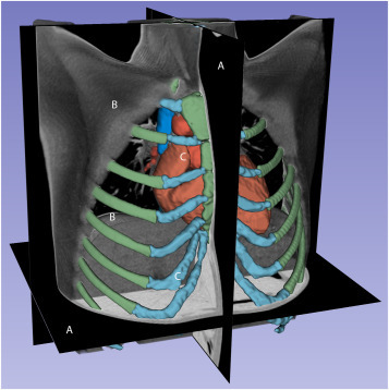
AI-generated 3D reconstructions provided accurate anatomical landmarks for preoperative planning in pediatric surgery, correlating closely with intraoperative anatomy. Key AI extensions - TotalSegmentator, MONAI Auto3DSeg, and RVesselX - ran locally, producing thoracic and abdominal segmentations in 60-90 seconds. Although trained on adult data, they reliably identified multiple pediatric structures for effective superimposition with volume-rendered images. Limitations included reduced accuracy in very young or low-weight patients and the lack of integrated intraoperative applications. Exported 3D models could also be used for education, VR/AR visualization, and 3D printing of surgical simulators or implants.
Making 3D segmentation accessible with AI
AI and computer vision have significantly lowered the technical and financial barriers to generating 3D reconstructions from standard CT and MRI scans. Publicly available tools now allow pediatric surgeons to create high-fidelity models quickly, without specialized expertise or expensive software. Future developments - such as AI models trained specifically on pediatric data - promise even greater accuracy and broader intraoperative and educational applications.
Medicalholodeck, combined with TotalSegmentator, makes segmentations available within minutes, providing fast, accessible 3D models without the need for manual processing. This significantly aids understanding of each patient's anatomy, enhances case planning, and may improve surgical outcomes.
For more information, contact info@medicalholodeck.com April 2026